Explore
Featured
Recent
Articles
Topics
Login
Upload
Featured
Recent
Articles
Topics
Login
Upload
Search Results for ''
published presentations and documents on DocSlides.
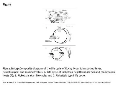
Figure Figure. Composite diagram of the life cycle of Rocky Mountain spotted fever, ricket
by willow
Azad AF, Beard CB. Rickettsial Pathogens and Their...
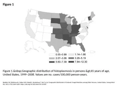
Figure 1 Figure 1. Geographic distribution of histoplasmosis in persons >65 years o
by davies
Baddley JW, Winthrop KL, Patkar NM, Delzell E, Beu...
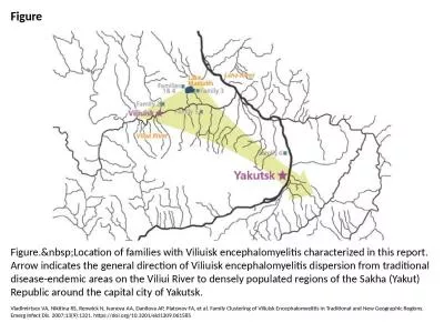
Figure Figure. Location of families with Viliuisk encephalomyelitis characterized in this
by grace3
Vladimirtsev VA, Nikitina RS, Renwick N, Ivanova A...
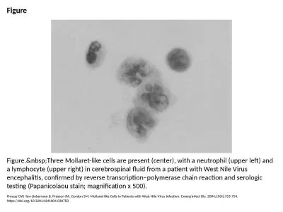
Figure Figure. Three Mollaret-like cells are present (center), with a neutrophil (upper le
by white
Procop GW, Yen-Lieberman B, Prayson RA, Gordon SM....
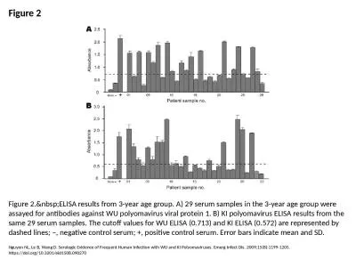
Figure 2 Figure 2. ELISA results from 3-year age group. A) 29 serum samples in the 3-year
by stella
Nguyen NL, Le B, Wang D. Serologic Evidence of Fre...
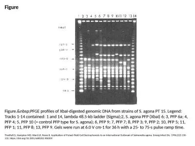
Figure Figure. PFGE profiles of XbaI-digested genomic DNA from strains of S. agona PT 15.
by megan
Threlfall EJ, Hampton MD, Ward LR, Rowe B. Applica...
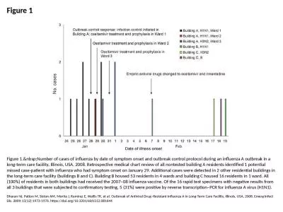
Figure 1 Figure 1. Number of cases of influenza by date of symptom onset and outbreak cont
by sophie
Dharan NJ, Patton M, Siston AM, Morita J, Ramirez ...
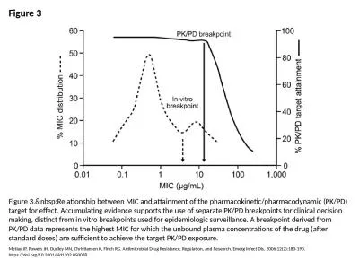
Figure 3 Figure 3. Relationship between MIC and attainment of the pharmacokinetic/pharmaco
by lucy
Metlay JP, Powers JH, Dudley MN, Christiansen K, F...
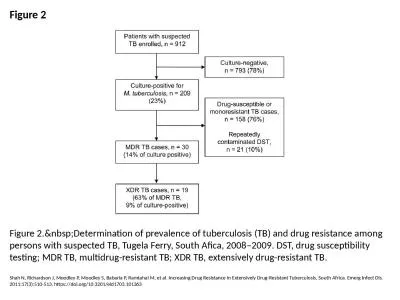
Figure 2 Figure 2. Determination of prevalence of tuberculosis (TB) and drug resistance am
by megan
Shah N, Richardson J, Moodley P, Moodley S, Babari...
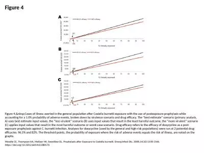
Figure 4 Figure 4. Cases of illness averted in the general population after Coxiella burne
by priscilla
Moodie CE, Thompson HA, Meltzer MI, Swerdlow DL. P...
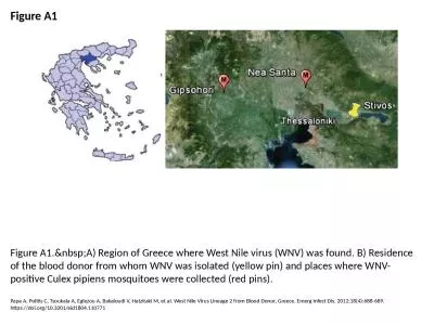
Figure A1 Figure A1. A) Region of Greece where West Nile virus (WNV) was found. B) Residen
by naomi
Papa A, Politis C, Tsoukala A, Eglezou A, Bakaloud...
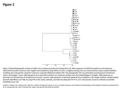
Figure 3 Figure 3. Phylogenetic analysis of nikB in IncI complex plasmids from Escherichia
by leah
Cottell JL, Webber MA, Coldham NG, Taylor DL, Cerd...
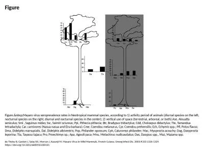
Figure Figure. Mayaro virus seroprevalence rates in Neotropical mammal species, according
by bethany
de Thoisy B, Gardon J, Salas RA, Morvan J, Kazanji...
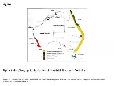
Figure Figure. Geographic distribution of rickettsial diseases in Australia.
by elyana
Odorico DM, Graves SR, Currie BJ, Catmull J, Nack ...
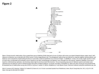
Figure 2 Figure 2. Genetic relationships of the spotted fever group rickettsiae (SFGR) det
by pagi
Silva N, Eremeeva ME, Rozental T, Ribeiro GS, Padd...
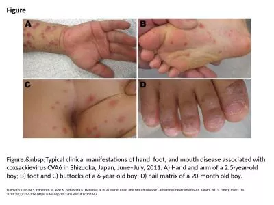
Figure Figure. Typical clinical manifestations of hand, foot, and mouth disease associated
by rose
Fujimoto T, Iizuka S, Enomoto M, Abe K, Yamashita ...
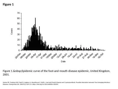
Figure 1 Figure 1. Epidemic curve of the foot-and-mouth disease epidemic, United Kingdom,
by gagnon
Hunter PR, Chalmers RM, Syed Q, Hughes LS, Woodhou...
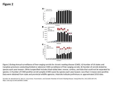
Figure 3 Figure 3. Distribution of PrPSc (disease-associated form of prion protein) in bra
by vivian
Angers RC, Seward TS, Napier D, Green M, Hoover E,...
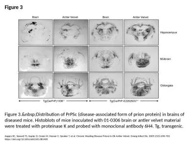
Figure 2 Figure 2. Annual surveillance of free-ranging cervids for chronic wasting disease
by roy
Saunders SE, Bartelt-Hunt SL, Bartz JC. Occurrence...
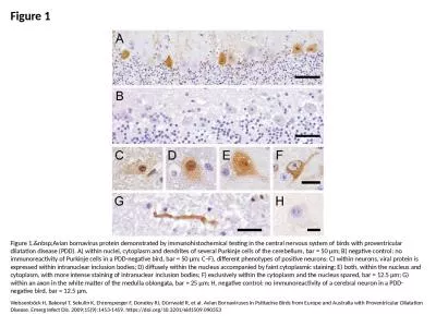
Figure 1 Figure 1. Avian bornavirus protein demonstrated by immunohistochemical testing in
by trinity
Weissenböck H, Bakonyi T, Sekulin K, Ehrensperger...
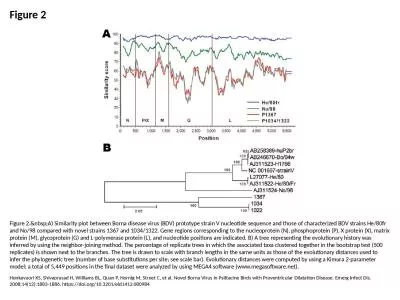
Figure 2 Figure 2. A) Similarity plot between Borna disease virus (BDV) prototype strain V
by dora
Honkavuori KS, Shivaprasad H, Williams BL, Quan P,...
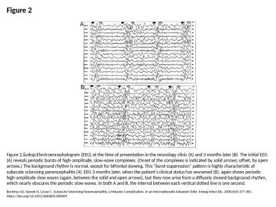
Figure 2 Figure 2. Electroencephalogram (EEG) at the time of presentation in the neurology
by finley
Bonthius DJ, Stanek N, Grose C. Subacute Sclerosin...
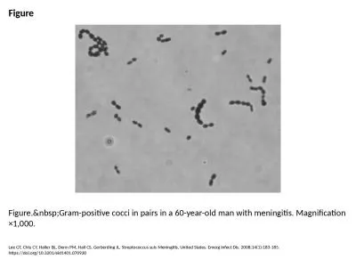
Figure Figure. Gram-positive cocci in pairs in a 60-year-old man with meningitis. Magnific
by bella
Lee GT, Chiu CY, Haller BL, Denn PM, Hall CS, Gerb...
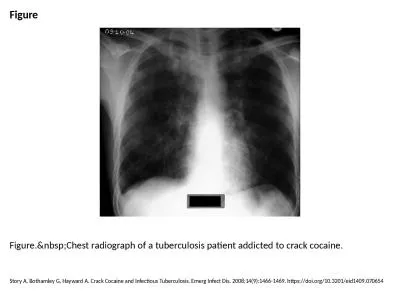
Figure Figure. Chest radiograph of a tuberculosis patient addicted to crack cocaine.
by mia
Story A, Bothamley G, Hayward A. Crack Cocaine and...
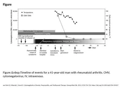
Figure Figure. Timeline of events for a 41-year-old man with rheumatoid arthritis. CMV, cy
by felicity
van Duin D, Miranda C, Husni E. Cytomegalovirus Vi...
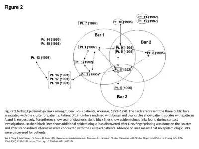
Figure 2 Figure 2. Epidemologic links among tuberculosis patients, Arkansas, 1992–1998.
by lauren
Ijaz K, Yang Z, Matthews HS, Bates JH, Cave MD. My...
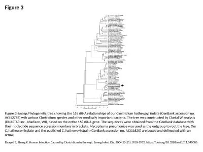
Figure 3 Figure 3. Phylogenetic tree showing the 16S rRNA relationships of our Clostridium
by rodriguez
Elsayed S, Zhang K. Human Infection Caused by Clos...
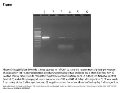
Figure Figure. Ethidium bromide stained agarose gel of ORF 1b standard reverse transcripti
by paige
Swayne DE, Suarez DL, Spackman E, Tumpey TM, Beck ...
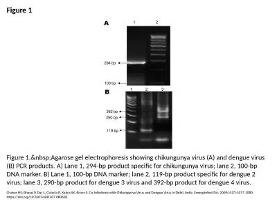
Figure 1 Figure 1. Agarose gel electrophoresis showing chikungunya virus (A) and dengue vi
by roy
Chahar HS, Bharaj P, Dar L, Guleria R, Kabra SK, B...
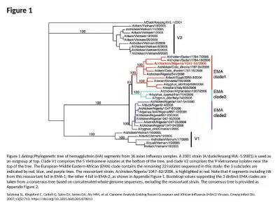
Figure 1 Figure 1. Phylogenetic tree of hemagglutinin (HA) segments from 36 avian influenz
by audrey
Salzberg SL, Kingsford C, Cattoli G, Spiro DJ, Jan...
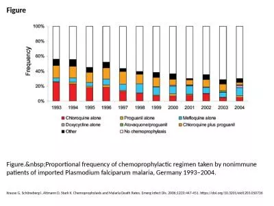
Figure Figure. Proportional frequency of chemoprophylactic regimen taken by nonimmune pati
by bery
Krause G, Schöneberg I, Altmann D, Stark K. Chemo...
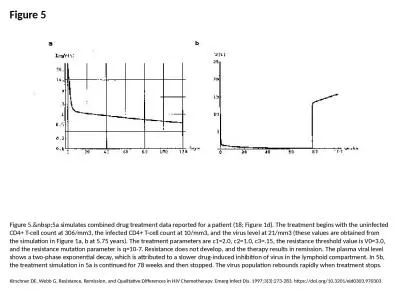
Figure 5 Figure 5. 5a simulates combined drug treatment data reported for a patient (18; F
by ximena
Kirschner DE, Webb G. Resistance, Remission, and Q...
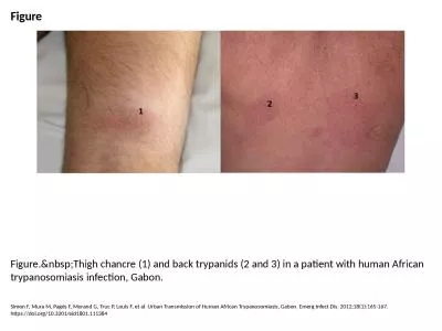
Figure Figure. Thigh chancre (1) and back trypanids (2 and 3) in a patient with human Afri
by martin
Simon F, Mura M, Pagès F, Morand G, Truc P, Louis...
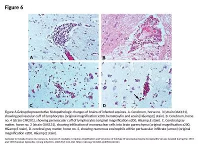
Figure 6 Figure 6. Representative histopathologic changes of brains of infected equines. A
by ida
Gonzalez D, Estrada-Franco JG, Carrara A, Aronson ...
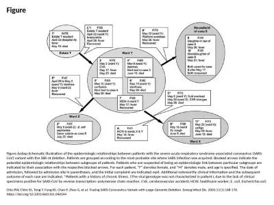
Figure Figure. Schematic illustration of the epidemiologic relationships between patients
by oconnor
Chiu RW, Chim SS, Tong Y, Fung KS, Chan P, Zhao G,...
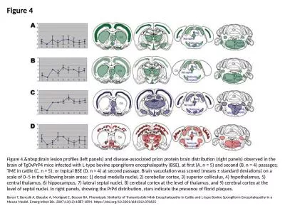
Figure 4 Figure 4. Brain lesion profiles (left panels) and disease-associated prion protei
by heavin
Baron T, Bencsik A, Biacabe A, Morignat E, Bessen ...
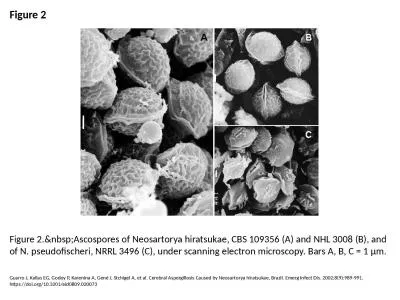
Figure 2 Figure 2. Ascospores of Neosartorya hiratsukae, CBS 109356 (A) and NHL 3008 (B),
by jainy
Guarro J, Kallas EG, Godoy P, Karenina A, Gené J,...
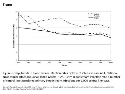
Figure Figure. Trends in bloodstream infection rates by type of intensive care unit, Natio
by mia
Gaynes R, Richards C, Edwards J, Emori TG, Horan T...
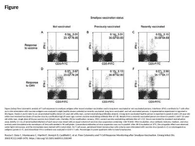
Figure Figure. Flow cytometric analysis of T-cell responses to smallpox antigens after rec
by patricia
Poccia F, Gioia C, Montesano C, Martini F, Horejsh...
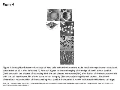
Figure 4 Figure 4. Atomic force microscopy of Vero cells infected with severe acute respir
by ella
Ng M, Lee J, Leong M, Ling A, Tan H, Ooi E. Topogr...
Load More...