Explore
Featured
Recent
Articles
Topics
Login
Upload
Featured
Recent
Articles
Topics
Login
Upload
Search Results for ''
published presentations and documents on DocSlides.
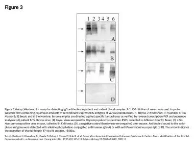
Figure 3 Figure 3. Western blot assay for detecting IgG antibodies in patient and rodent b
by naomi
Torrez-Martinez N, Bharadwaj M, Goade D, Delury J,...
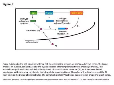
Figure 3 Figure 3. Cell-to-cell signaling systems. Cell-to-cell signaling systems are comp
by pagi
Van Delden C, Iglewski BH. Cell-to-Cell Signaling ...
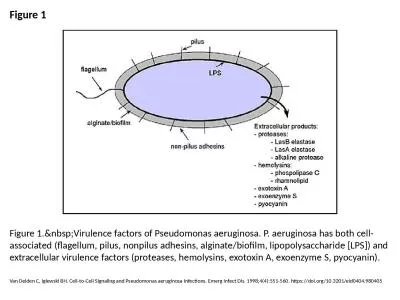
Figure 1 Figure 1. Virulence factors of Pseudomonas aeruginosa. P. aeruginosa has both cel
by lydia
Van Delden C, Iglewski BH. Cell-to-Cell Signaling ...
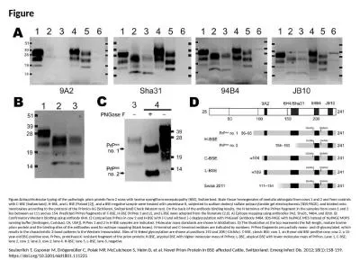
Figure Figure. Molecular typing of the pathologic prion protein from 2 cows with bovine sp
by rose
Seuberlich T, Gsponer M, Drögemüller C, Polak MP...
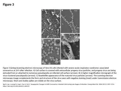
Figure 3 Figure 3. Scanning electron microscopy of Vero E6 cells infected with severe acut
by madeline
Ng M, Lee J, Leong M, Ling A, Tan H, Ooi E. Topogr...
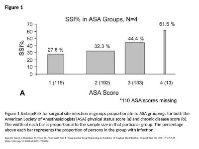
Figure 1 Figure 1. Risk for surgical site infection in groups proportionate to ASA groupin
by violet
Kaye KS, Sands K, Donahue JG, Chan KA, Fishman P, ...
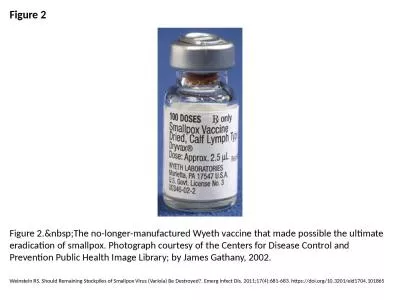
Figure 2 Figure 2. The no-longer-manufactured Wyeth vaccine that made possible the ultimat
by caroline
Weinstein RS. Should Remaining Stockpiles of Small...
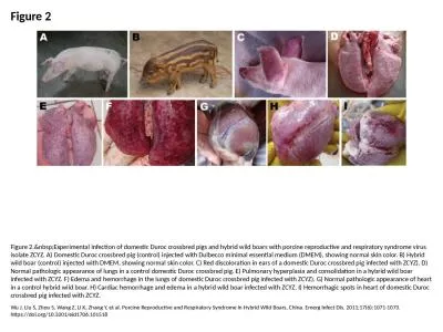
Figure 2 Figure 2. Experimental infection of domestic Duroc crossbred pigs and hybrid wild
by ava
Wu J, Liu S, Zhou S, Wang Z, Li K, Zhang Y, et al....
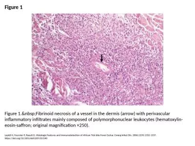
Figure 1 Figure 1. Fibrinoid necrosis of a vessel in the dermis (arrow) with perivascular
by catherine
Lepidi H, Fournier P, Raoult D. Histologic Feature...
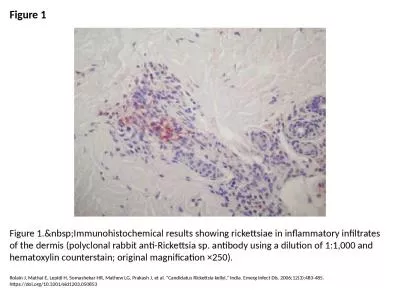
Figure 1 Figure 1. Immunohistochemical results showing rickettsiae in inflammatory infiltr
by roy
Rolain J, Mathai E, Lepidi H, Somashekar HR, Mathe...
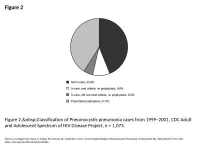
Figure 2 Figure 2. Classification of Pneumocystis pneumonia cases from 1999–2001, CDC Ad
by ruby
Morris A, Lundgren JD, Masur H, Walzer PD, Hanson ...
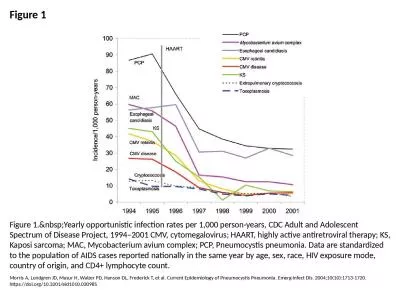
Figure 1 Figure 1. Yearly opportunistic infection rates per 1,000 person-years, CDC Adult
by sophia2
Morris A, Lundgren JD, Masur H, Walzer PD, Hanson ...
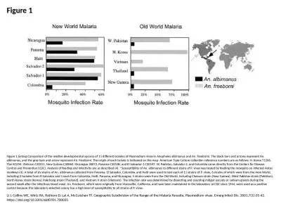
Figure 1 Figure 1. Comparison of the relative developmental success of 11 different isolat
by byrne
Li J, Collins WE, Wirtz RA, Rathore D, Lal A, McCu...
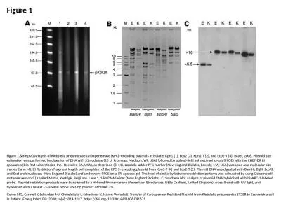
Figure 1 Figure 1. A) Analysis of Klebsiella pneumoniae carbapenemase (KPC)–encoding pla
by bitsy
Goren MG, Carmeli Y, Schwaber MJ, Chmelnitsky I, S...
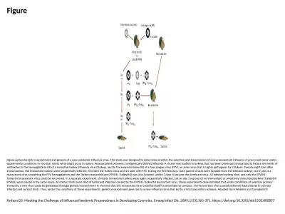
Figure Figure. Genetic reassortment and genesis of a new pandemic influenza virus. This st
by bery
Fedson DS. Meeting the Challenge of Influenza Pand...
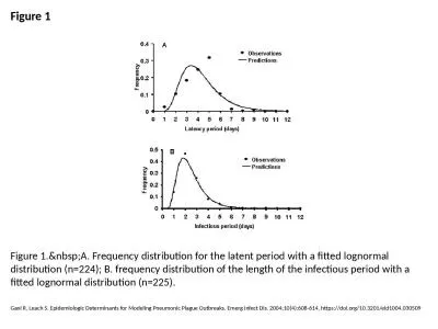
Figure 1 Figure 1. A. Frequency distribution for the latent period with a fitted lognormal
by ethlyn
Gani R, Leach S. Epidemiologic Determinants for Mo...
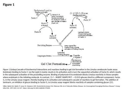
Figure 1 Figure 1. Cascade of biochemical interactions and reactions leading to gel clot f
by molly
Miller-Hjelle MA, Hjelle JT, Jones M, Mayberry WR,...
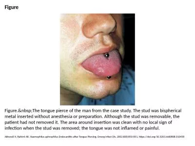
Figure Figure. The tongue pierce of the man from the case study. The stud was bispherical
by cecilia
Akhondi H, Rahimi AR. Haemophilus aphrophilus Endo...
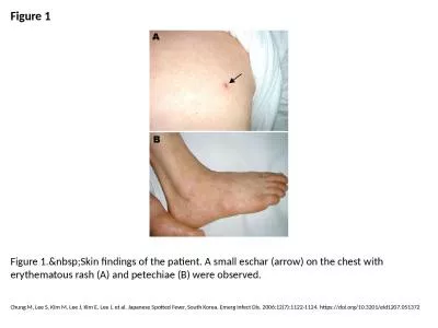
Figure 1 Figure 1. Skin findings of the patient. A small eschar (arrow) on the chest with
by eloise
Chung M, Lee S, Kim M, Lee J, Kim E, Lee J, et al....
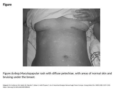
Figure Figure. Maculopapular rash with diffuse petechiae, with areas of normal skin and br
by nicole
Delgado MJ, Gutierrez JM, Radic LB, Maretic T, Zek...
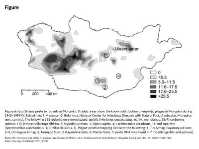
Figure Figure. Yersina pestis in rodents in Mongolia. Shaded areas show the known distribu
by violet
Riehm JM, Tserennorov D, Kiefer D, Stuermer IW, To...
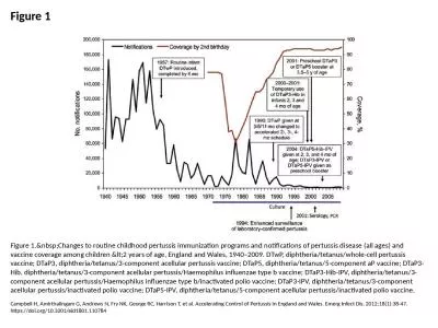
Figure 1 Figure 1. Changes to routine childhood pertussis immunization programs and notifi
by bency
Campbell H, Amirthalingam G, Andrews N, Fry NK, Ge...
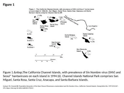
Figure 1 Figure 1. The California Channel Islands, with prevalence of Sin Nombre virus (SN
by susan
Graham TB, Chomel BB. Population Dynamics of the D...
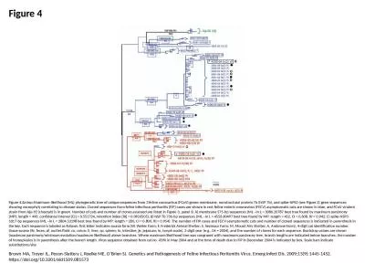
Figure 4 Figure 4. Maximum-likelihood (ML) phylogenetic tree of unique sequences from 3 fe
by elise
Brown MA, Troyer JL, Pecon-Slattery J, Roelke ME, ...
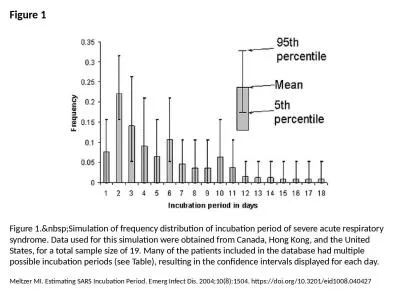
Figure 1 Figure 1. Simulation of frequency distribution of incubation period of severe acu
by jalin
Meltzer MI. Estimating SARS Incubation Period. Eme...
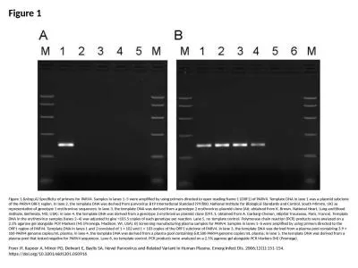
Figure 1 Figure 1. A) Specificity of primers for PARV4. Samples in lanes 1–5 were amplif
by berey
Fryer JF, Kapoor A, Minor PD, Delwart E, Baylis SA...
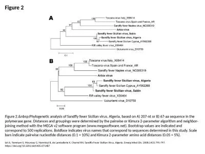
Figure 2 Figure 2. Phylogenetic analysis of Sandfly fever Sicilian virus, Algeria, based o
by naomi
Izri A, Temmam S, Moureau G, Hamrioui B, de Lambal...
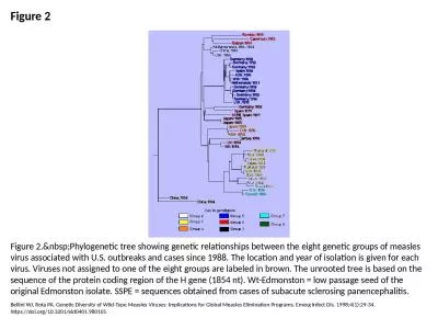
Figure 2 Figure 2. Phylogenetic tree showing genetic relationships between the eight genet
by clara
Bellini WJ, Rota PA. Genetic Diversity of Wild-Typ...
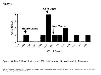
Figure 1 Figure 1. Epidemiologic curve of Yersinia enterocolitica outbreak in Tennessee.
by unita
Jones TF, Buckingham SC, Bopp CA, Ribot E, Schaffn...
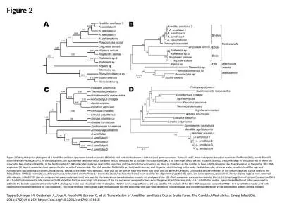
Figure 2 Figure 2. Molecular phylogeny of 4 Armillifer amillatus specimens based on partia
by dandy
Tappe D, Meyer M, Oesterlein A, Jaye A, Frosch M, ...
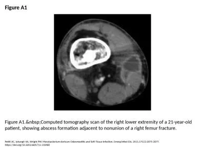
Figure A1 Figure A1. Computed tomography scan of the right lower extremity of a 21-year-ol
by melody
Pettit AC, Jahangir AA, Wright PW. Mycobacterium d...
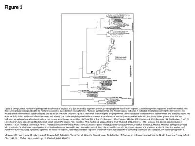
Figure 1 Figure 1. Overall hantavirus phylogenetic tree based on analysis of a 139 nucleot
by emmy
Monroe MC, Morzunov SP, Johnson AM, Bowen MD, Arts...
Figure 1 Figure 1. Chest radiographs of patient T/Tx at hospital admission on August 20, 1
by hanah
Torrez-Martinez N, Bharadwaj M, Goade D, Delury J,...
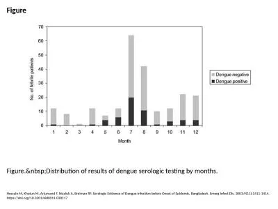
Figure Figure. Distribution of results of dengue serologic testing by months.
by pamela
Hossain M, Khatun M, Arjumand F, Nisaluk A, Breima...
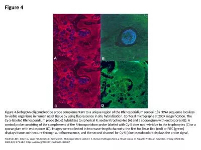
Figure 4 Figure 4. An oligonucleotide probe complementary to a unique region of the Rhinos
by jovita
Fredricks DN, Jolley JA, Lepp PW, Kosek JC, Relman...
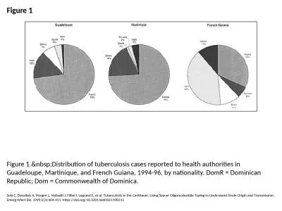
Figure 1 Figure 1. Distribution of tuberculosis cases reported to health authorities in Gu
by tracy
Sola C, Devallois A, Horgen L, Maïsetti J, Fillio...
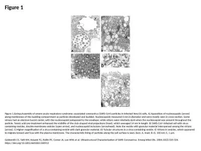
Figure 1 Figure 1. Assembly of severe acute respiratory syndrome–associated coronavirus
by edolie
Goldsmith CS, Tatti KM, Ksiazek TG, Rollin PE, Com...
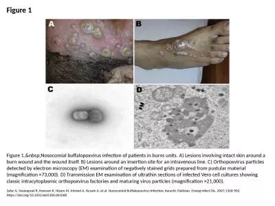
Figure 1 Figure 1. Nosocomial buffalopoxvirus infection of patients in burns units. A) Les
by lauren
Zafar A, Swanepoel R, Hewson R, Nizam M, Ahmed A, ...
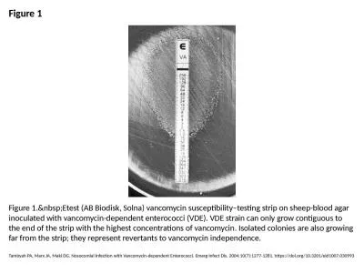
Figure 1 Figure 1. Etest (AB Biodisk, Solna) vancomycin susceptibility–testing strip on
by ruby
Tambyah PA, Marx JA, Maki DG. Nosocomial Infection...
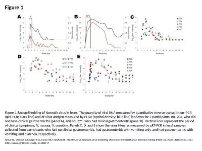
Figure 1 Figure 1. Shedding of Norwalk virus in feces. The quantity of viral RNA measured
by brooke
Atmar RL, Opekun AR, Gilger MA, Estes MK, Crawford...
Load More...