Explore
Featured
Recent
Articles
Topics
Login
Upload
Featured
Recent
Articles
Topics
Login
Upload
Search Results for 'electron microscopy'
electron microscopy published presentations and documents on DocSlides.
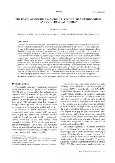
Microscopical studies on horse eosinophils73Braz J morphol Sci 20
by stella
THE HORSE EOSINOPHIL AS A MODEL LEUCOCYTE FOR MORP...
Microscopes and Cells 2.1.4
by alexa-scheidler
Comparison of relative sizes of molecules, cell m...
SEM & TEM in Polymer Characterization
by cheryl-pisano
Scanning Electron Microscopy (SEM). Uses. Sample ...
Characterizing epitaxially
by danika-pritchard
-grown . InGaAs. quantum dot chains using transm...
Center for Nanoscale Materials
by adah
Building 440 Phone: 630 - 252 - 1775 Fax: 630 - 25...
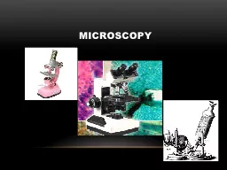
Microscopy Microscope DEFINE:
by sophie
instrument that produces . enlarged . image of . o...
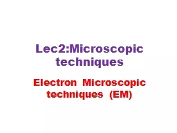
Lec2:Microscopic techniques
by zoe
Electron . Microscopic techniques . (EM). 2020-202...
Downloaded from httprupressorgjcbarticlepdf262669138334266
by genevieve
Downloaded from http://rupress.org/jcb/article-pdf...
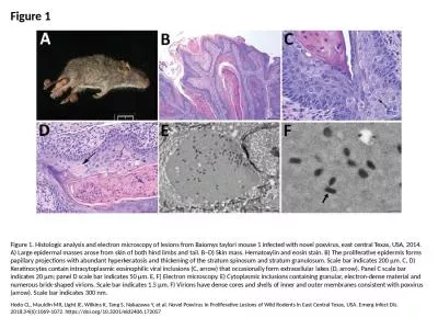
Figure 1 Figure 1. Histologic analysis and electron microscopy of lesions from Baiomys taylori mous
by cora
Hodo CL, Mauldin MR, Light JE, Wilkins K, Tang S, ...
Year 8 Lesson 8 – cells
by delilah
Science. Learning intention. To know how microscop...
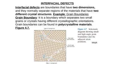
INTERFACIAL DEFECTS Interfacial defects
by emma
are boundaries that have . two dimensions,. and t...
Electron Microscopy Laboratory
by bency
Introduction to transmission electron microscopy. ...
1 Microscopy and
by phoebe-click
Specimen Preparation. Microscope. Basics...
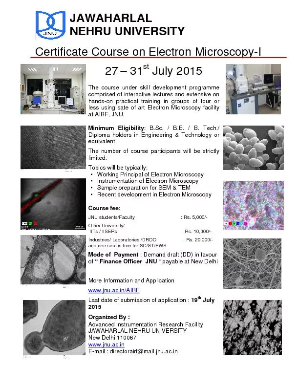
JAWAHARLAL NEHRU UNIVERSITY Certificate Course on Electron Microscopy
by briana-ranney
Microscopy and Magnification
by marina-yarberry
1000. 1000. 1000. 1000. mm. Micrometre. n. m. Nan...
Automation in Single-Particle Electron Microscopy
by natalia-silvester
Jian. Guan. Hafenstein Lab. The definition of Au...
Failure of an Inconel 718 die used in production of hot cop
by danika-pritchard
M. Schwartz, D. Gheorghe, R. . Ciocoiu. , I. . Ci...
Scanning Electron Microscopy (SEM)
by pasty-toler
. . Conclusions. Changin...
Quasi-liquid layers on ice crystal surfaces
by tatyana-admore
Sazaki. , et al., 2012: . Quasi-liquid layers on ...
Electron Microscopy and You in the New
by alexa-scheidler
M. illennium. The Challenges in Electron Microsco...
Occupational
by liane-varnes
Exposure Assessments of Nanomaterials in the USA....
CORRELATIVE VIDEO-LIGHT ELECTRON MICROSCOPY In studies of dynamic cel
by pamella-moone
This module describes a newly developed method tha...
Failure of an Inconel 718 die used in production of hot cop
by yoshiko-marsland
M. Schwartz, D. Gheorghe, R. . Ciocoiu. , I. . Ci...
Fluorescence and Scanning Electron Microscopy of ChitosanDNA Nanoparticles for Biological Applications A
by olivia-moreira
Masotti F Marino G Ortagg i and Cleofe Palocci D...
Electron Tomography a Powerful Tool for D Cellular Microscopy Technical renements enable investigators to locate proteins inside cells and to anticipate tomographic reconstructions of bacteria Sriram
by yoshiko-marsland
In the last 64257ve decades electron microscopy E...
Cryo-Electron Microscopy
by alexa-scheidler
James Conway. University of Pittsburgh School of ...
The Microscope and Cell Theory
by min-jolicoeur
The Microscope. An understanding of cells and the...
Investigation of the microstructure in the Yttrium-
by aaron
Tantalate. -. Zirconia. system. Thursday, August...
Developments in
by morgan
Reel-to-Reel Electron MicroscopyInfrastructureChri...
Microscopes & Stains
by lucinda
تقنية الأجهزة . الكيموحيوية...
muscle fibers Ingels 1997 The papillary muscle and thethe ventricu
by riley
papillary muscle by scanning electron microscopy,M...
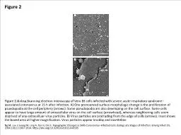
Figure 2 Figure 2. Scanning electron microscopy of Vero E6 cells infected with severe acut
by clara
Ng M, Lee J, Leong M, Ling A, Tan H, Ooi E. Topogr...
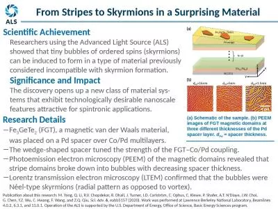
Research Details Fe 3 GeTe
by cadie
2. (FGT), a magnetic van der Waals material, . wa...
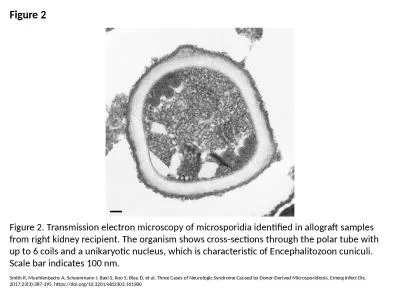
Figure 2 Figure 2. Transmission electron microscopy of microsporidia identified in allograft sample
by rodriguez
Smith R, Muehlenbachs A, Schaenmann J, Baxi S, Koo...
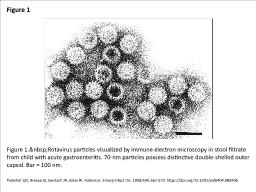
Figure 1 Figure 1. Rotavirus particles visualized by immune electron microscopy in stool f
by olivia
Parashar UD, Bresee JS, Gentsch JR, Glass RI. Rota...
An Introduction to Electron Microscopy
by julia
Because these electron micrograph (EM) images are ...
Evaluation of a Prototype, Triple-Tube, Water-based Nano Spot-Collector: Application to Microscopic
by hailey
Presenter: Orthodoxia Zervaki. Authors: Orthodoxia...
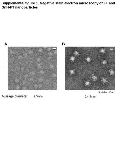
Supplemental figure 1. Negative stain electron microscopy of FT and GnH-FT nanoparticles
by morgan
Scale bar: 10nm. 9.5nm. 14.7nm. Average diameter:....
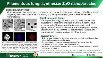
Filamentous fungi synthesize ZnO nanoparticles
by genevieve
Scientific Achievement. We demonstrate that bioche...
ISAC CASE STUDY SOOT-IN-OIL DIAGNOSTICS
by jacey
ISAC_CS_08. Soot-in-Oil Diagnostics. Transmission ...
Load More...