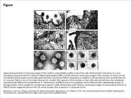
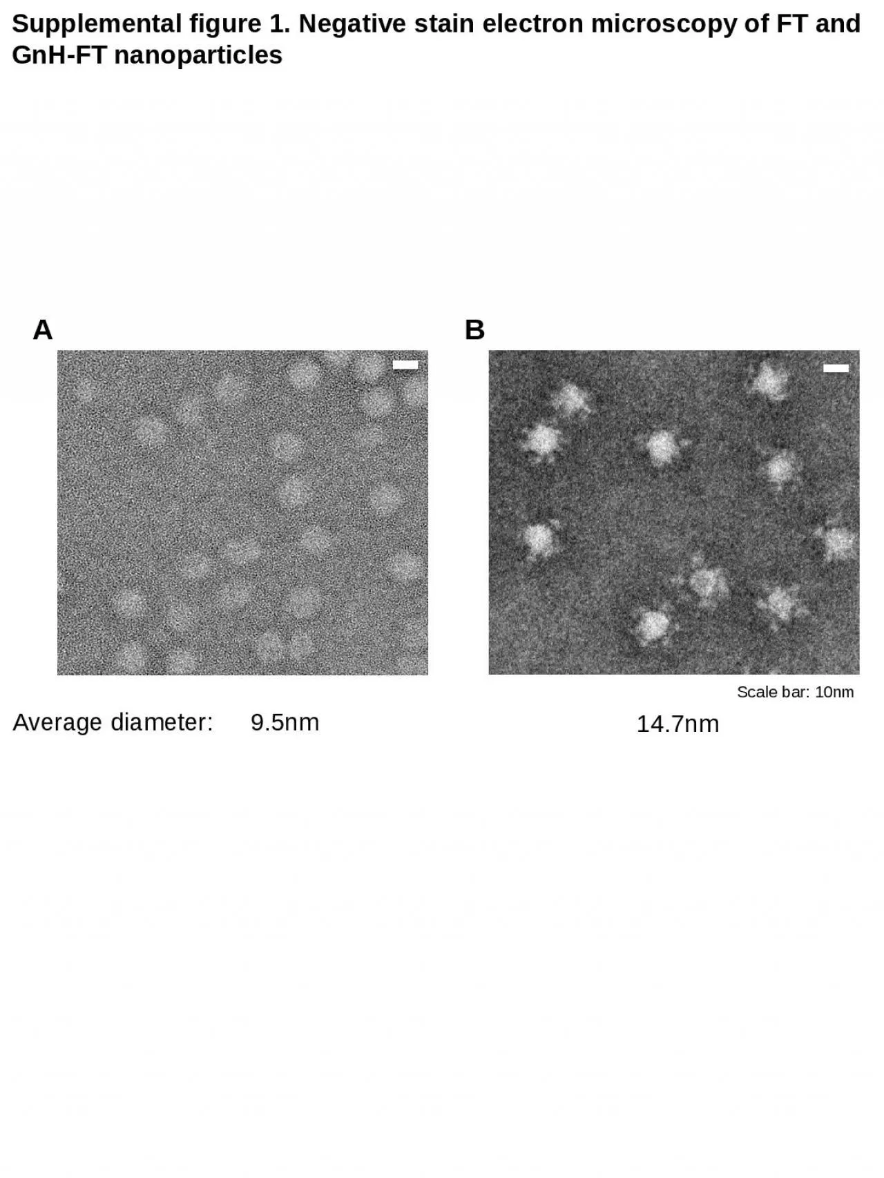
PPT-Supplemental figure 1. Negative stain electron microscopy of FT and GnH-FT nanoparticles

Author : morgan | Published Date : 2024-01-03

Scale bar 10nm 95nm 147nm Average diameter A B Supplemental figure 2 ELISA using FT and GnHFT as antigens A B P lt005 Plt001 Plt0001 and Plt000001

Presentation Embed Code

Download Presentation

Download Presentation The PPT/PDF document "Supplemental figure 1. Negative stain el..." is the property of its rightful owner. Permission is granted to download and print the materials on this website for personal, non-commercial use only, and to display it on your personal computer provided you do not modify the materials and that you retain all copyright notices contained in the materials. By downloading content from our website, you accept the terms of this agreement.

Supplemental figure 1. Negative stain electron microscopy of FT and GnH-FT nanoparticles: Transcript


Scale bar 10nm 95nm 147nm Average diameter A B Supplemental figure 2 ELISA using FT and GnHFT as antigens A B P lt005 Plt001 Plt0001 and Plt000001 . Chelsea Aitken. Peter Aspinall. http://www.zyvexlabs.com/EIPBNuG/EIPBN%20images/05Ion.jpg. Intro to Electron Microscopy. Similar to optical microscopy except with electrons rather than photons. Used to image samples with a resolution of 10 Å. M. illennium. The Challenges in Electron Microscopy Concerning Negative Stain and . CryoEM. Techniques for Virology . Reseach. . With a General Review of The Baker Lab Paper Too. Bob Ashley AAS SMFW. chool . curriculums are . influenced . by the dominant . worldview driven by market force. The ability to create greed is what success in business is defined . I. ndigenous . source of knowledge and wisdom, and more importantly the indigenous . Supplemental Figure 1 Supplemental Figure 2 Supplemental Figure 3 Supplemental Figure 4 Supplemental Table 1 Supplemental Figure 5 Replica of van 1670 Moody Use the information in this tutorial to supplement the visuals in lab and the Chapters 1 8 and 9 in your lab manual Replica of Culpepper tripod microscope built c 1725 by Co DMSs . and dietary intake and lifestyle factors at . exam 5 . for . all participants (n=1880). . -log10 (P value). 60. 50. 40. 30. 20. 10. 132 Nutrients/Bioactive . 129 Food group1 (FFD). 29 Food group2 (FG5Serv). tissues, cells, cell components, cell contents, or microorganisms.. *The stain may be . natural or synthetic. . . *The object stained is called the . substrate. . . *Staining facilitates the observation of a substrate by introducing . (SEM) Electron Microscopy (SEM) and TEM Scanning electron microscopy is used for inspecting topographies of specimens at very high magnifications using a piece of equipment called the scanning electr The theory of image formation in electron microscope s is very similar to image formation in light microscope s. However, the practice and design are completely different : The most crucial Riquelme C, Escors D, Ortego J, Sanchez CM, Uzelac-Keserovic B, Apostolov K, et al. Nature of the Virus Associated with Endemic Balkan Nephropathy. Emerg Infect Dis. 2002;8(8):869-870. https://doi.org/10.3201/eid0808.020042. O'Rourke LG, Pitulle C, Hegarty BC, Kraycirik S, Killary KA, Grosenstein P, et al. Bartonella quintana in Cynomolgus Monkey (Macaca fascicularis). Emerg Infect Dis. 2005;11(12):1931-1934. https://doi.org/10.3201/eid1112.030045. The aims of this course are:. ● . to introduce the principles of scanning electron microscopy. ● . to describe the components of the microscope and explain how they work. ● . to highlight some of the problems which can arise during imaging. suboptimally. positioned. There was a normal . cardiothymic. silhouette. The lungs were clear. Air in scattered loops of . nondilated. small and large bowel, within all four quadrants of the abdomen.. ELECTRON. . MICROSC. O. PY. . . . Electron. . micros. c. opy. . (EM). . is a technique. . u. s. ed. . for. . obtaining. . high re. s. olu. t. ion. . images. . of. . biological and non-biologi.

Download Document

Here is the link to download the presentation.
"Supplemental figure 1. Negative stain electron microscopy of FT and GnH-FT nanoparticles"The content belongs to its owner. You may download and print it for personal use, without modification, and keep all copyright notices. By downloading, you agree to these terms.

Related Documents