Search Results for 'amp figure'
amp figure published presentations and documents on DocSlides.
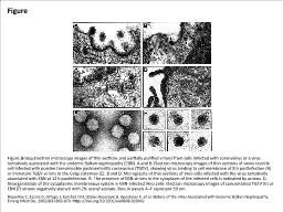
Figure Figure. Electron microscopy images of thin sections and partially purified virions
by faith
Riquelme C, Escors D, Ortego J, Sanchez CM, Uzelac...
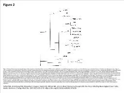
Figure 2 Figure 2. Maximum parsimony phylogram indicating the relationship of deer tick vi
by ash
Telford SR, Armstrong PM, Katavolos P, Foppa I, Ga...
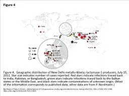
Figure 4 Figure 4. Geographic distribution of New Delhi metallo-β-lactamase-1 producers,
by megan
Nordmann P, Naas T, Poirel L. Global Spread of Car...
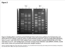
Figure 2 Figure 2. XbaI and NotI pulsed-field gel electrophoresis patterns for Escherichia
by riley
Vincent C, Boerlin P, Daignault D, Dozois CM, Duti...
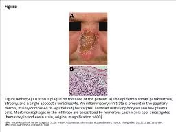
Figure Figure. A) Crustosus plaque on the nose of the patient. B) The epidermis shows para
by audrey
Faber WR, Hoekzema R, Bart A, Zeegelaar JE, de Vri...
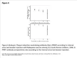
Figure 4 Figure 4. A. Plaque-reduction neutralizing antibody titers (PRNT) according to in
by miller
Sohn YM. Japanese Encephalitis Immunization in Sou...
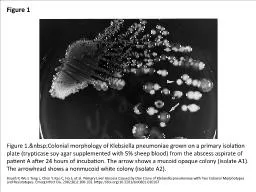
Figure 1 Figure 1. Colonial morphology of Klebsiella pneumoniae grown on a primary isolati
by badra
Hsueh P, Wu J, Teng L, Chen Y, Kao C, Ho S, et al....
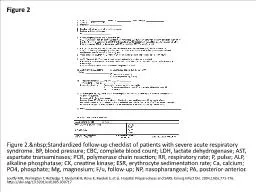
Figure 2 Figure 2. Standardized follow-up checklist of patients with severe acute respirat
by alyssa
Loutfy MR, Wallington T, Rutledge T, Mederski B, R...
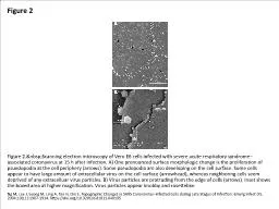
Figure 2 Figure 2. Scanning electron microscopy of Vero E6 cells infected with severe acut
by clara
Ng M, Lee J, Leong M, Ling A, Tan H, Ooi E. Topogr...
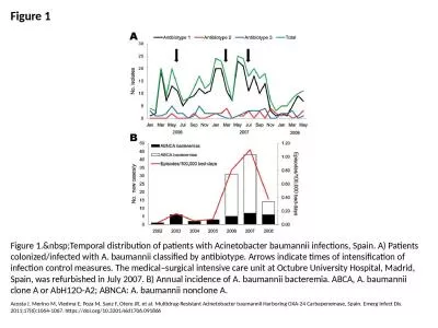
Figure 1 Figure 1. Temporal distribution of patients with Acinetobacter baumannii infectio
by taylor
Acosta J, Merino M, Viedma E, Poza M, Sanz F, Oter...
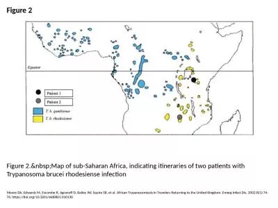
Figure 2 Figure 2. Map of sub-Saharan Africa, indicating itineraries of two patients with
by eve
Moore DA, Edwards M, Escombe R, Agranoff D, Bailey...
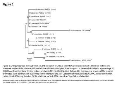
Figure 1 Figure 1. Neighbor-joining tree of a 1,341-bp region of unique 16S rRNA gene sequ
by lydia
Simmon KE, Brown-Elliott BA, Ridge PG, Durtschi JD...
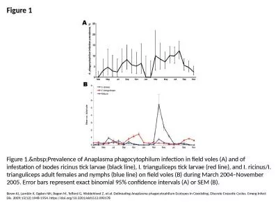
Figure 1 Figure 1. Prevalence of Anaplasma phagocytophilum infection in field voles (A) an
by ceila
Bown KJ, Lambin X, Ogden NH, Begon M, Telford G, W...
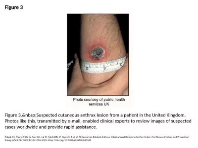
Figure 3 Figure 3. Suspected cutaneous anthrax lesion from a patient in the United Kingdom
by megan
Polyak CS, Macy JT, De La Cruz MI, Lai JE, McAulif...
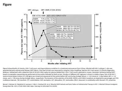
Figure Figure. Kinetics of viremia, CD4+ T-cell count, and drug resistance mutations in a
by amber
Bansal V, Metzner KJ, Niederöst B, Leemann C, Bö...
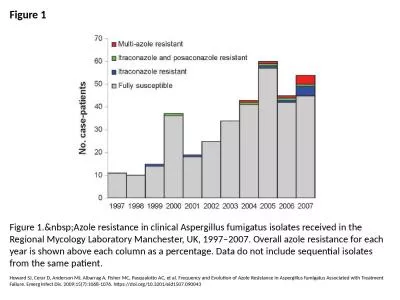
Figure 1 Figure 1. Azole resistance in clinical Aspergillus fumigatus isolates received in
by smith
Howard SJ, Cerar D, Anderson MJ, Albarrag A, Fishe...
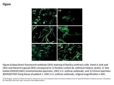
Figure Figure. Direct fluorescent-antibody (DFA) staining of Bacillus anthracis cells. Pan
by ruby
De BK, Bragg SL, Sanden GN, Wilson KE, Diem LA, Ma...
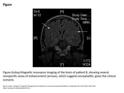
Figure Figure. Magnetic resonance imaging of the brain of patient B, showing several nonsp
by angelina
Kee AC, Yang S, Tambyah P. Atypical Chikungunya Vi...
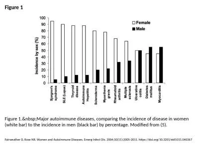
Figure 1 Figure 1. Major autoimmune diseases, comparing the incidence of disease in women
by bitsy
Fairweather D, Rose NR. Women and Autoimmune Disea...
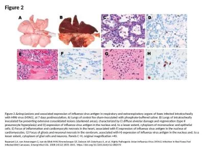
Figure 2 Figure 2. Lesions and associated expression of influenza virus antigen in respira
by joy
Reperant LA, van Amerongen G, van de Bildt MW, Rim...
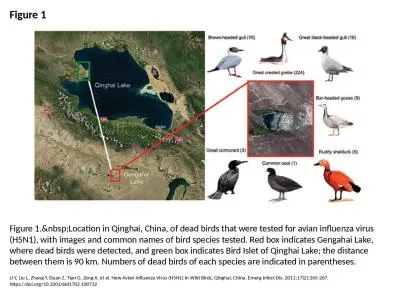
Figure 1 Figure 1. Location in Qinghai, China, of dead birds that were tested for avian in
by smith
Li Y, Liu L, Zhang Y, Duan Z, Tian G, Zeng X, et a...
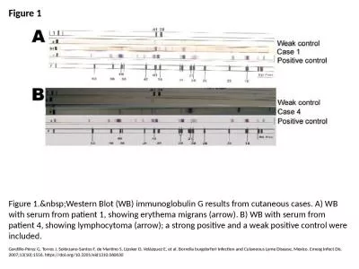
Figure 1 Figure 1. Western Blot (WB) immunoglobulin G results from cutaneous cases. A) WB
by ida
Gordillo-Pérez G, Torres J, Solórzano-Santos F, ...
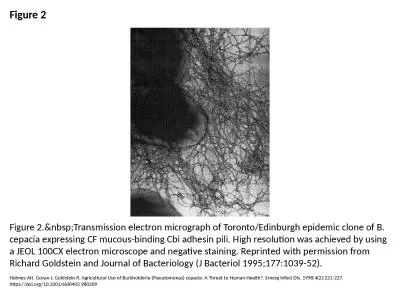
Figure 2 Figure 2. Transmission electron micrograph of Toronto/Edinburgh epidemic clone of
by danya
Holmes AH, Govan J, Goldstein R. Agricultural Use ...
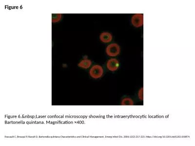
Figure 6 Figure 6. Laser confocal microscopy showing the intraerythrocytic location of Bar
by hadly
Foucault C, Brouqui P, Raoult D. Bartonella quinta...
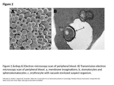
Figure 2 Figure 2. A) Electron microscopy scan of peripheral blood. B) Transmission electr
by emma
O'Rourke LG, Pitulle C, Hegarty BC, Kraycirik S, K...
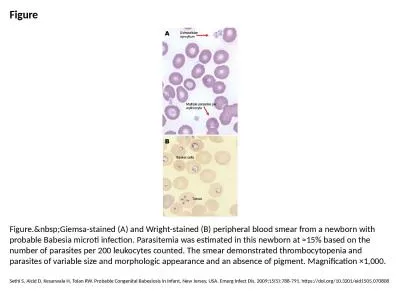
Figure Figure. Giemsa-stained (A) and Wright-stained (B) peripheral blood smear from a new
by deena
Sethi S, Alcid D, Kesarwala H, Tolan RW. Probable ...

Figure 1 Figure 1. Lesions of the patient infected with Babesia divergens 1 day after hosp
by samantha
Haapasalo K, Suomalainen P, Sukura A, Siikamäki H...
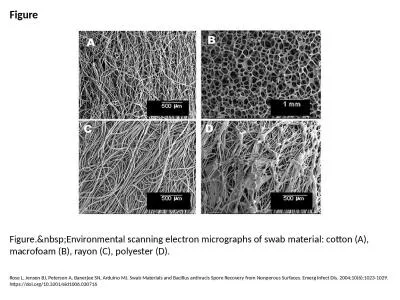
Figure Figure. Environmental scanning electron micrographs of swab material: cotton (A), m
by cecilia
Rose L, Jensen BJ, Peterson A, Banerjee SN, Arduin...
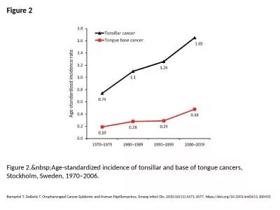
Figure 2 Figure 2. Age-standardized incidence of tonsillar and base of tongue cancers, Sto
by wang
Ramqvist T, Dalianis T. Oropharyngeal Cancer Epide...
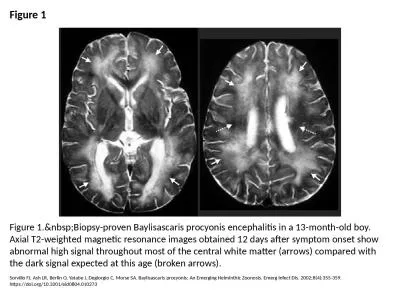
Figure 1 Figure 1. Biopsy-proven Baylisascaris procyonis encephalitis in a 13-month-old bo
by susan
Sorvillo FJ, Ash LR, Berlin O, Yatabe J, Degiorgio...
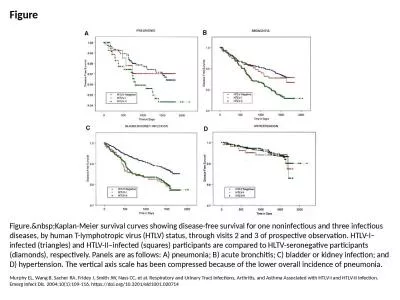
Figure Figure. Kaplan-Meier survival curves showing disease-free survival for one noninfec
by daisy
Murphy EL, Wang B, Sacher RA, Fridey J, Smith JW, ...
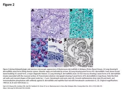
Figure 2 Figure 2. Histopathologic and electron microscopic appearance of Blastomyces derm
by oneill
Harris JR, Blaney DD, Lindsley MD, Zaki SR, Paddoc...
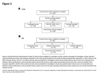
Figure 2 Figure 2. Flowchart displaying the number of blood cultures eligible for evaluati
by abigail
Trick WE, Zagorski BM, Tokars JI, Vernon MO, Welbe...
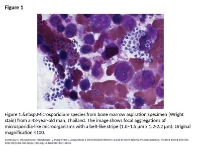
Figure 1 Figure 1. Microsporidium species from bone marrow aspiration specimen (Wright sta
by leah
Suankratay C, Thiansukhon E, Nilaratanakul V, Puta...
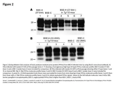
Figure 2 Figure 2. Western blot analyses of brain protease-resistant prion protein (PrPres
by kimberly
Torres J, Andréoletti O, Lacroux C, Prieto I, Lor...
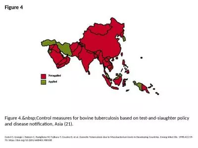
Figure 4 Figure 4. Control measures for bovine tuberculosis based on test-and-slaughter po
by smith
Cosivi O, Grange J, Daborn C, Raviglione M, Fujiku...
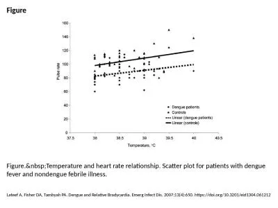
Figure Figure. Temperature and heart rate relationship. Scatter plot for patients with den
by jordyn
Lateef A, Fisher DA, Tambyah PA. Dengue and Relati...
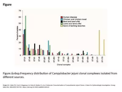
Figure Figure. Frequency distribution of Campylobacter jejuni clonal complexes isolated fr
by eloise
Dingle KE, Colles FM, Ure R, Wagenaar JA, Duim B, ...
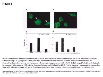
Figure 4 Figure 4. Mitochondria transmembrane potential and caspase activities measurement
by reese
Yang J, Ramanathan MP, Muthumani K, Choo AY, Jin S...
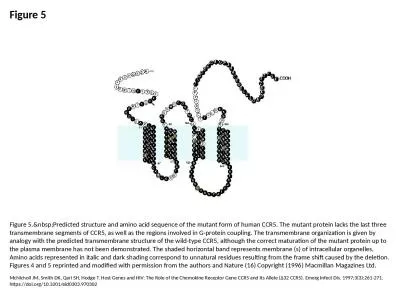
Figure 5 Figure 5. Predicted structure and amino acid sequence of the mutant form of human
by candy
McNicholl JM, Smith DK, Qari SH, Hodge T. Host Gen...