Explore
Featured
Recent
Articles
Topics
Login
Upload
Featured
Recent
Articles
Topics
Login
Upload
Search Results for ''
published presentations and documents on DocSlides.
In a radiograph of average density , the radiographic images of dental structures can be divided
by arya
into 2 . groups. . Anatomical Landmarks ...
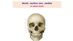
Maxilla , maxillary sinus,
by melody
mandible. Dr. . Gallatz. Katalin. Parts. of . ...
Dental Radiographic Positioning
by delcy
Stephen . Juriga. DVM, Dipl. AVDC. www.veterinary...
Cysts of the jaws Cysts may occur centrally (within bone) in any location in the maxilla or mandibl
by hadly
Odontogenic. cysts are found most often in the to...
Fractures of the middle and lower third of the faces.
by bella
BMS L. ECTURES . SERIES. MANDIBULAR FRACTURES. Sec...
Parotid Region [ para =around,
by fanny
otic. =ear]. Presented by :- Dr. . Sushma. . Tom...
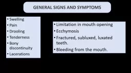
GENERAL SIGNS AND SYMPTOMS
by elena
Swelling. Pain. Drooling. Tenderness . Bony discon...
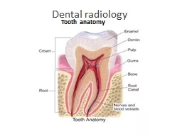
Dental radiology Tooth anatomy
by arya
Crown. The visible part of the tooth above the gum...
Odontogenic tumors Introduction
by hadley
Tooth formation is a complex process that involves...
The Axial Sekelton Skull
by mila-milly
Divisions of the Skeleton. The skeleton is divided...
ROLE OF MULTIDETECTOR SPIRAL-CT IN THE MANAGEMENT OF MAXILLO-FACIAL
by taylor
TRAUMATIC LESIONS. Department of Neuroradiology. ....
Load More...