Explore
Featured
Recent
Articles
Topics
Login
Upload
Featured
Recent
Articles
Topics
Login
Upload
Search Results for 'image cells'
image cells published presentations and documents on DocSlides.
USER GUIDETali ImageBased CytometerCatalog Number Publication Number
by belinda
x0000x0000 x/MCIxD 0 x/MCIxD 0 Information in this...
Tali Viability Kit 150 Dead Cell Greenfor use with Tali Assays Green G
by cecilia
MaterialConcentrationStorageStabilityTali Dead Cel...
Tali Viability Kit – Dead Cell Green*for use with Tali Assays: Gr
by beatrice
MaterialConcentrationStorage*StabilityTali Dead Ce...
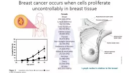
Breast cancer occurs when cells proliferate uncontrollably in breast tissue
by olivia-moreira
BRCA2 is a tumor suppressor gene responsible for ...
The Human Eye 14.2 – Pages 449-455
by stefany-barnette
The Human Eye 14.2 – Pages 449-455 A) Types of ...
Quantitative Phase Imaging of Cells and Tissues
by lindy-dunigan
Chaps.1-2. Introduction and groundwork. Light Mic...
S patial Analysis: Raster
by marina-yarberry
Rasters. are . beautiful.. Rasters. don’t dep...
Any guesses as to what I research at U of A?
by faustina-dinatale
Mr. Zach Dean, M.S.. Fluorescent Probes for Cance...
The Illusion of Mental Pictures
by briana-ranney
Zenon Pylyshyn. Rutgers University,. Center for ...
Finish Batteries
by sherrill-nordquist
Review Capacity. Peukert's. Equation - . I = cu...
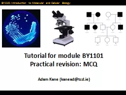
Tutorial for module BY1101
by lindy-dunigan
Practical revision: MCQ. Adam Kane (kanead@tcd.ie...
An Introduction to
by luanne-stotts
Electron Microscopy. Because these electron micro...
Microscope
by giovanna-bartolotta
Instrument used to make very small objects appear...
ARTIFICIAL EYE BATCH C2 Vibhor
by dorothy
. jain. . Mainak. . chakraborty. Rohan. . seth....
Photonics West February 3, 2013 Brian C. Smith, Ph.D. ,Princeton Instruments Jason McClure, Ph.D. Princeton Instruments Dan Heller, Ph.D. Memorial Sloan-Kettering Cancer Center Ed Gooding, Ph.D. Princeton Instruments
by everett673
Brian C. Smith, Ph.D. ,Princeton Instruments . Jas...
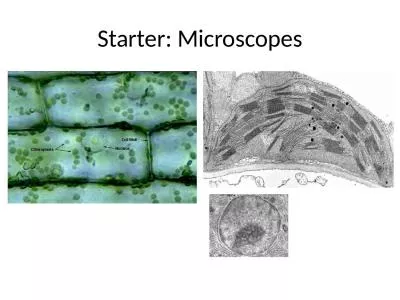
Starter: Microscopes Block 1A - Cell structure 2.1.1
by joyce
Microscopy. Foundations in Biology. SPEC. Objectiv...
Year 8 Lesson 8 – cells
by delilah
Science. Learning intention. To know how microscop...
Creating Colors Part 3: Cones, color, curves, and afterimages!
by murphy
New Hampshire Destination Imagination • nh-di.or...
NERVOUS SYSTEM SPECIAL SENSES
by osullivan
SPECIAL SNESES. Those which are not somatic senses...
Lesson 5 : The Human Eye
by elizabeth
SCIENCE 8 UNIT 2: Optics. YOUR TASKS:. Take out y...
An Introduction to Electron Microscopy
by julia
Because these electron micrograph (EM) images are ...
Games and Animation Occlusion Culling
by melanie
CSE 3541. Prof. Roger Crawfis. Culling Problem. Ha...
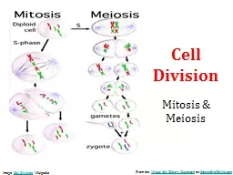
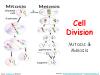
Cell Division Mitosis & Meiosis
by melody
From the . Virtual Cell Biology Classroom. on . ...
GASTROINTESTINAL MOTILITY
by tabitha
PHYSIOLOGY JAYA PUNATI, MD DIRECTOR, PEDIATRIC G...
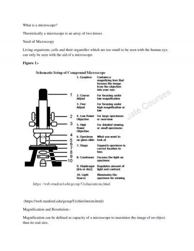
What is a microscope
by elizabeth
Theoretically a microscope is an array of two lens...
Virtual Cell Biology Classroom
by bethany
Cell Division Mitosis & Meiosis From the on S...
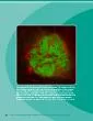
Mouse pancreatic islet imaged with confocal microscopy This image rev
by anderson
THE BETA CELL Adult Mouse Cells Reprogrammed To B...
Combining High Throughput Screening with Image-Based Phenotyping
by WiseWhale
to advance APBD . drug . discovery. Or Kakhlon . D...
The “Science for Dummies”
by PlayfulPenguin
of a Pastoral Encounter. Karen Elsworth. The Biolo...
Utility of t2wi in differentiating gct and chordoma of sacrum
by victoria
. - Dr. Ayesha . Erum. Hadi. . MD- . Radiodi...
Load More...