Explore
Featured
Recent
Articles
Topics
Login
Upload
Featured
Recent
Articles
Topics
Login
Upload
Search Results for 'Figure-Inches'
Figure-Inches published presentations and documents on DocSlides.
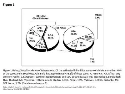
Figure 1 Figure 1. Global incidence of tuberculosis. Of the estimated 8.8 million cases wo
by callie
Rattan A, Kalia A, Ahmad N. Multidrug-Resistant My...
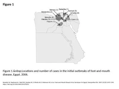
Figure 1 Figure 1. Locations and number of cases in the initial outbreaks of foot-and-mout
by reese
Knowles NJ, Wadsworth J, Reid SM, Swabey KG, El-Kh...
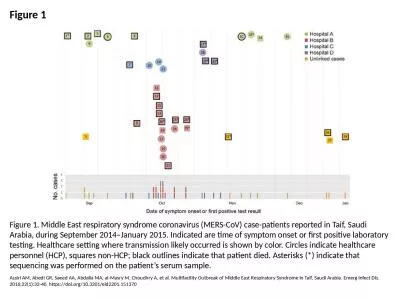
Figure 1 Figure 1. Middle East respiratory syndrome coronavirus (MERS-CoV) case-patients reported i
by brown
Assiri AM, Abedi GR, Saeed AA, Abdalla MA, al-Masr...
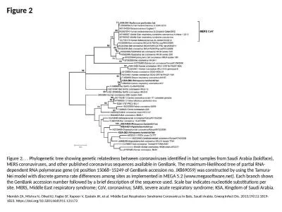
Figure 2 Figure 2. . . Phylogenetic tree showing genetic relatedness between coronaviruses identifi
by cady
Memish ZA, Mishra N, Olival KJ, Fagbo SF, Kapoor V...
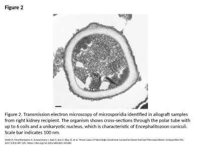
Figure 2 Figure 2. Transmission electron microscopy of microsporidia identified in allograft sample
by rodriguez
Smith R, Muehlenbachs A, Schaenmann J, Baxi S, Koo...
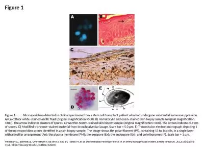
Figure 1 Figure 1. . . . . Microsporidium detected in clinical specimens from a stem cell transplan
by isabella2
Meissner EG, Bennett JE, Qvarnstrom Y, da Silva A,...
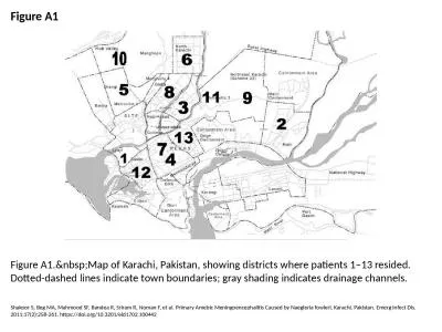
Figure A1 Figure A1. Map of Karachi, Pakistan, showing districts where patients 1–13 res
by elizabeth
Shakoor S, Beg MA, Mahmood SF, Bandea R, Sriram R,...
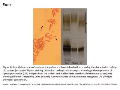
Figure Figure. A) Gram stain of pus from the patient’s submental collection, showing the
by taylor
Brent AJ, Matthews PC, Dance DA, Pitt TL, Handy R....
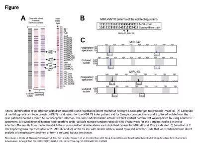
Figure Figure. Identification of co-infection with drug-susceptible and reactivated latent multidru
by brooke
Pérez-Lago L, Lirola M, Navarro Y, Herranz M, Rui...
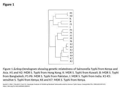
Figure 1 Figure 1. Dendogram showing genetic relatedness of Salmonella Typhi from Kenya an
by roxanne
Kariuki S, Gilks C, Revathi G, Hart CA. Genotypic ...
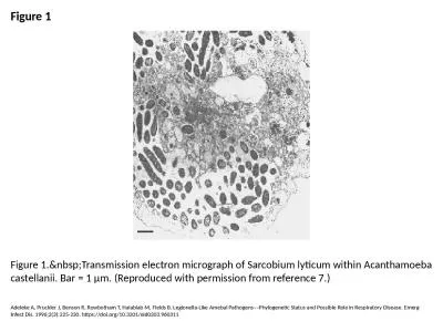
Figure 1 Figure 1. Transmission electron micrograph of Sarcobium lyticum within Acanthamoe
by elise
Adeleke A, Pruckler J, Benson R, Rowbotham T, Hala...
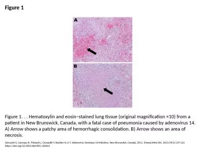
Figure 1 Figure 1. . . Hematoxylin and eosin–stained lung tissue (original magnification ×10) fr
by elina
Girouard G, Garceau R, Thibault L, Oussedik Y, Bas...
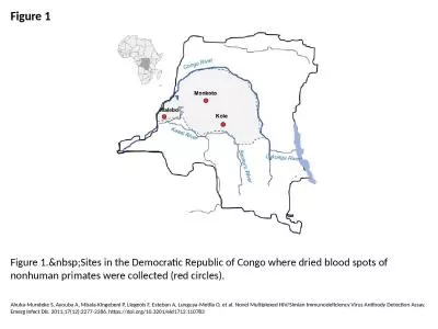
Figure 1 Figure 1. Sites in the Democratic Republic of Congo where dried blood spots of no
by cora
Ahuka-Mundeke S, Ayouba A, Mbala-Kingebeni P, Lieg...
Figure 2 Figure 2. Identification of diverse Simian immunodeficiency virus (SIV) lineages
by barbara
Peeters M, Courgnaud V, Abela B, Auzel P, Pourrut ...
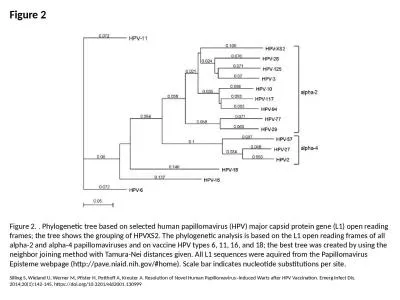
Figure 2 Figure 2. . Phylogenetic tree based on selected human papillomavirus (HPV) major capsid pr
by esther
Silling S, Wieland U, Werner M, Pfister H, Potthof...
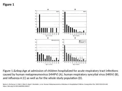
Figure 1 Figure 1. Age at admission of children hospitalized for acute respiratory tract i
by cappi
Boivin G, De Serres G, Côté S, Gilca R, Abed Y, ...
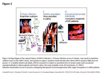
Figure 2 Figure 2. Stages of the natural history of HHV-6 infection: I. Primary infection
by olivia
Campadelli-Fiume G, Mirandola P, Menotti L. Human ...
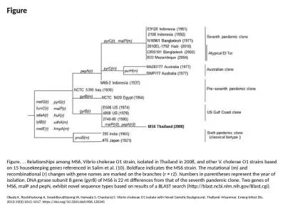
Figure Figure. . . Relationships among MS6, Vibrio cholerae O1 strain, isolated in Thailand in 2008
by vivian
Okada K, Roobthaisong A, Swaddiwudhipong W, Hamada...
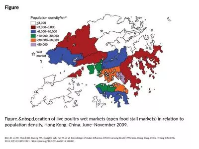
Figure Figure. Location of live poultry wet markets (open food stall markets) in relation
by priscilla
Kim JH, Lo FK, Cheuk KK, Kwong MS, Goggins WB, Cai...
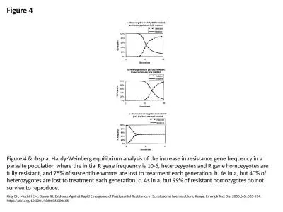
Figure 4 Figure 4. a. Hardy-Weinberg equilibrium analysis of the increase in resistance ge
by dora
King CH, Muchiri EM, Ouma JH. Evidence Against Rap...
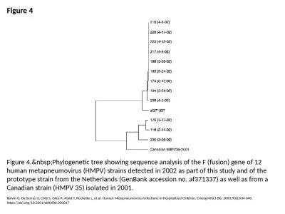
Figure 4 Figure 4. Phylogenetic tree showing sequence analysis of the F (fusion) gene of 1
by cecilia
Boivin G, De Serres G, Côté S, Gilca R, Abed Y, ...
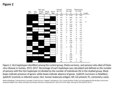
Figure 2 Figure 2. HLA haplotypes identified among the control group, Ebola survivors, and persons
by rodriguez
Wawina-Bokalanga T, Vanmechelen B, Lhermitte V, Ma...
Figure Figure. Chest radiograph of patient who acquired acute pulmonary histoplasmosis aft
by piper
Weinberg M, Weeks J, Lance-Parker S, Traeger M, Wi...
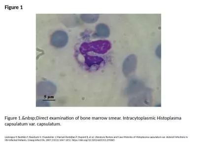
Figure 1 Figure 1. Direct examination of bone marrow smear. Intracytoplasmic Histoplasma c
by adah
Loulergue P, Bastides F, Baudouin V, Chandenier J,...
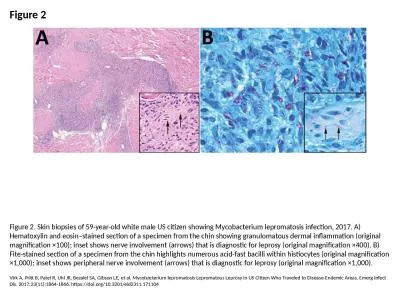
Figure 2 Figure 2. Skin biopsies of 59-year-old white male US citizen showing Mycobacterium leproma
by olivia
Virk A, Pritt B, Patel R, Uhl JR, Bezalel SA, Gibs...
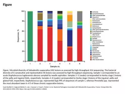
Figure Figure. Microbial diversity of hidradenitis suppurativa (HS) lesions as assessed by high-thr
by davies
Guet-Revillet H, Coignard-Biehler H, Jais J, Quesn...
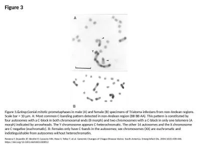
Figure 3 Figure 3. Gonial mitotic prometaphases in male (A) and female (B) specimens of Tr
by garcia
Panzera F, Dujardin JP, Nicolini P, Caraccio MN, R...
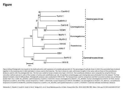
Figure Figure. Phylogenetic tree based on the deduced amino acid sequences of complete gly
by grace3
Watanabe S, Maeda K, Suzuki K, Ueda N, Iha K, Tani...
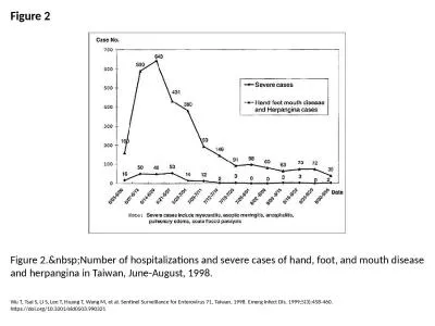
Figure 2 Figure 2. Number of hospitalizations and severe cases of hand, foot, and mouth di
by adia
Wu T, Tsai S, Li S, Lee T, Huang T, Wang M, et al....
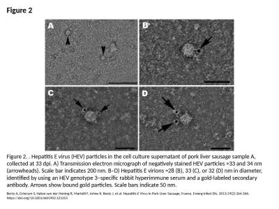
Figure 2 Figure 2. . Hepatitis E virus (HEV) particles in the cell culture supernatant of pork live
by freya
Berto A, Grierson S, Hakze-van der Honing R, Marte...
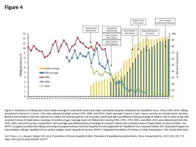
Figure 4 Figure 4. Prevalence of HBsAg and 3-dose HepB coverage for each birth cohort and major vac
by wilson
Cui F, Shen L, Li L, Wang H, Wang F, Bi S, et al. ...
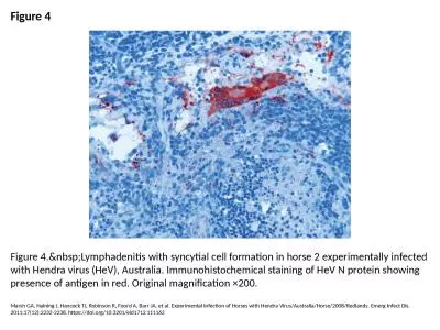
Figure 4 Figure 4. Lymphadenitis with syncytial cell formation in horse 2 experimentally i
by trinity
Marsh GA, Haining J, Hancock TJ, Robinson R, Foord...
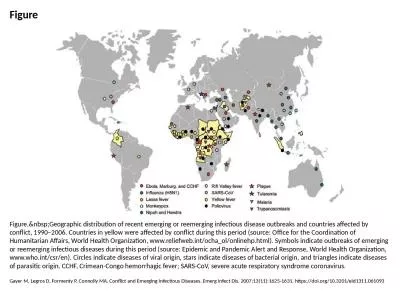
Figure Figure. Geographic distribution of recent emerging or reemerging infectious disease
by anderson
Gayer M, Legros D, Formenty P, Connolly MA. Confli...
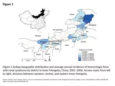
Figure 1 Figure 1. Geographic distribution and average annual incidence of hemorrhagic fev
by helene
Zhang Y, Zhang F, Gao N, Wang J, Zhao Z, Li M, et ...
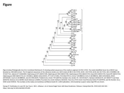
Figure Figure. Phylogenetic tree (CLC-Combined Workbench 3) showing partial sequences of t
by jordyn
Huong VT, Yoshimatsu K, Luan VD, Van Tuan L, Nhi L...
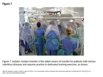
Figure 7 Figure 7. Isolator–isolator transfer is the safest means of transfer for patients with s
by julia
Nicol ED, Mepham S, Naylor J, Mollan I, Adam M, d�...
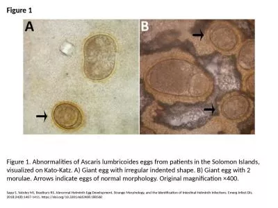
Figure 1 Figure 1. Abnormalities of Ascaris lumbricoides eggs from patients in the Solomon Islands,
by angelina
Sapp S, Yabsley MJ, Bradbury RS. Abnormal Helminth...
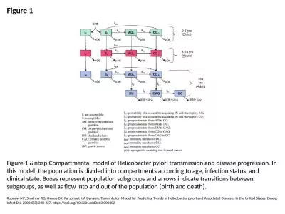
Figure 1 Figure 1. Compartmental model of Helicobacter pylori transmission and disease pro
by kylie
Rupnow MF, Shachter RD, Owens DK, Parsonnet J. A D...
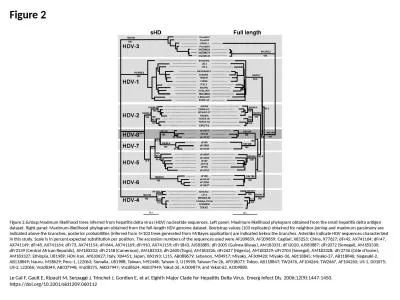
Figure 2 Figure 2. Maximum likelihood trees inferred from hepatitis delta virus (HDV) nucl
by josephine
Le Gal F, Gault E, Ripault M, Serpaggi J, Trinchet...
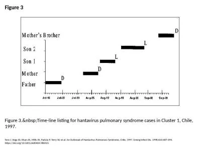
Figure 3 Figure 3. Time-line listing for hantavirus pulmonary syndrome cases in Cluster 1,
by jaena
Toro J, Vega JD, Khan AS, Mills JN, Padula P, Terr...
Load More...