Explore
Featured
Recent
Articles
Topics
Login
Upload
Featured
Recent
Articles
Topics
Login
Upload
Search Results for 'Figure-Hedgehog'
Figure-Hedgehog published presentations and documents on DocSlides.
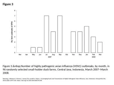
Figure 3 Figure 3. Number of highly pathogenic avian influenza (H5N1) outbreaks, by month,
by melody
Henning J, Wibawa H, Morton J, Usman TB, Junaidi A...
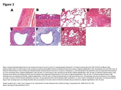
Figure 3 Figure 3. Histopathologic lesions in the trachea and lungs of control (A and C) o
by maisie
Song D, Kang B, Lee C, Saif LJ, Ha G, Kang D, et a...
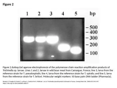
Figure 2 Figure 2. Gel agarose electrophoresis of the polymerase chain reaction amplificat
by bella
Ranque S, Faugère B, Pozio E, La Rosa G, Tamburri...
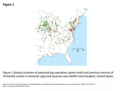
Figure 1 Figure 1. Locations of pastured-pig operations (green dots) and previous records
by hanah
Burke R, Masuoka P, Murrell KD. Swine Trichinella ...
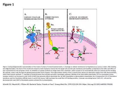
Figure 1 Figure 1. Diagrammatic representation of the mode of action of several bacterial
by isla
Schmitt CK, Meysick KC, O'Brien AD. Bacterial Toxi...
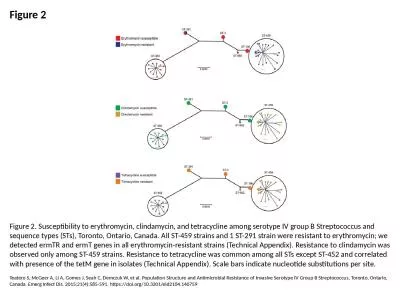
Figure 2 Figure 2. Susceptibility to erythromycin, clindamycin, and tetracycline among serotype IV
by mackenzie
Teatero S, McGeer A, Li A, Gomes J, Seah C, Demczu...
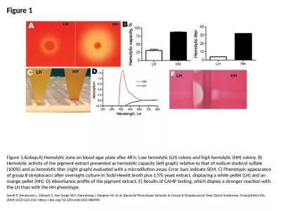
Figure 1 Figure 1. A) Hemolytic zone on blood agar plate after 48 h: Low hemolytic (LH) co
by mia
Sendi P, Johansson L, Dahesh S, Van Sorge NM, Dare...
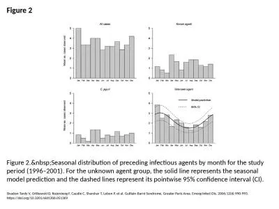
Figure 2 Figure 2. Seasonal distribution of preceding infectious agents by month for the s
by roxanne
Sivadon-Tardy V, Orlikowski D, Rozenberg F, Caudie...
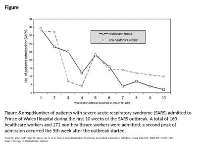
Figure Figure. Number of patients with severe acute respiratory syndrome (SARS) admitted t
by roxanne
Chan PK, Ip M, Ng K, Chan RC, Wu A, Lee N, et al. ...
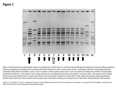
Figure 1 Figure 1. Molecular epidemiology of group A streptococcus (GAS) strains in outbre
by jade
Smith A, Li A, Tolomeo O, Tyrell GJ, Jamieson FB, ...
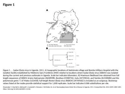
Figure 1 Figure 1. . . Sudan Ebola virus in Uganda, 2011. A) Geographic locations of Nakisimata vil
by tremblay
Shoemaker T, MacNeil A, Balinandi S, Campbell S, W...
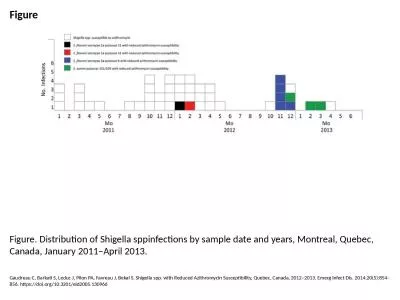
Figure Figure. Distribution of Shigella sppinfections by sample date and years, Montreal, Quebec, C
by bella
Gaudreau C, Barkati S, Leduc J, Pilon PA, Favreau ...
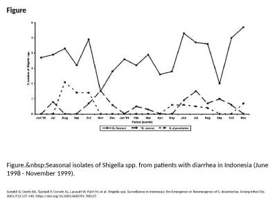
Figure Figure. Seasonal isolates of Shigella spp. from patients with diarrhea in Indonesia
by udeline
Subekti D, Oyofo BA, Tjaniadi P, Corwin AL, Larasa...
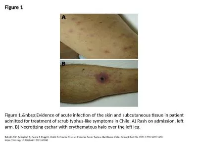
Figure 1 Figure 1. Evidence of acute infection of the skin and subcutaneous tissue in pati
by patricia
Balcells ME, Rabagliati R, García P, Poggi H, Odd...
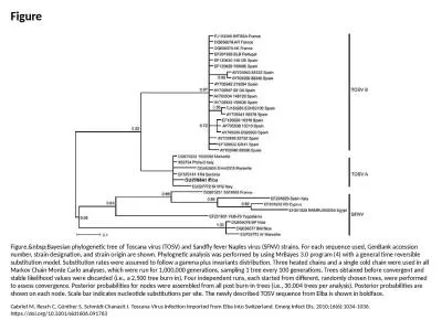
Figure Figure. Bayesian phylogenetic tree of Toscana virus (TOSV) and Sandfly fever Naples
by harmony
Gabriel M, Resch C, Günther S, Schmidt-Chanasit J...
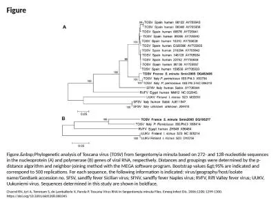
Figure Figure. Phylogenetic analysis of Toscana virus (TOSV) from Sergentomyia minuta base
by pagi
Charrel RN, Izri A, Temmam S, de Lamballerie X, Pa...
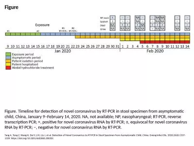
Figure Figure. Timeline for detection of novel coronavirus by RT-PCR in stool specimen from asympto
by claire
Tang A, Tong Z, Wang H, Dai Y, Li K, Liu J, et al....
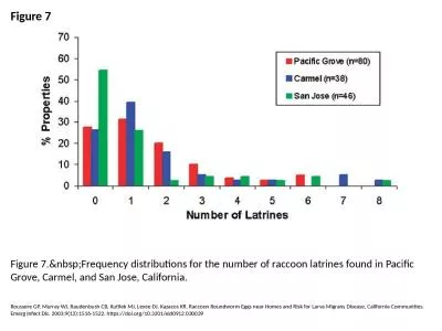
Figure 7 Figure 7. Frequency distributions for the number of raccoon latrines found in Pac
by reese
Roussere GP, Murray WJ, Raudenbush CB, Kutilek MJ,...
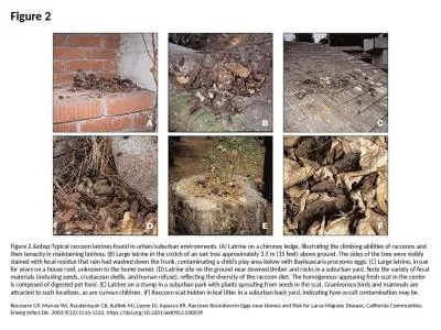
Figure 2 Figure 2. Typical raccoon latrines found in urban/suburban environments. (A) Latr
by sophie
Roussere GP, Murray WJ, Raudenbush CB, Kutilek MJ,...
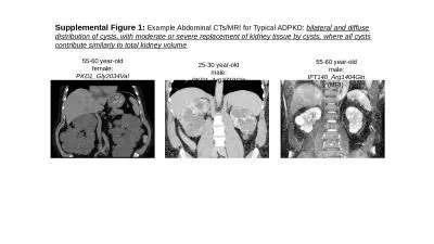
Supplemental Figure 1: Example Abdominal CTs/MRI for Typical ADPKD:
by helene
bilateral and diffuse distribution of cysts, with ...
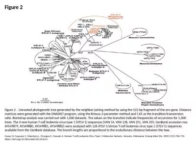
Figure 2 Figure 2. . Unrooted phylogenetic tree generated by the neighbor-joining method by using t
by hazel
Cassar O, Capuano C, Meertens L, Chungue E, Gessai...
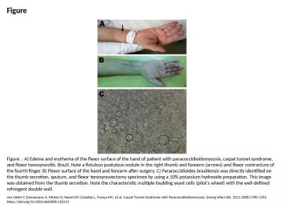
Figure Figure. . A) Edema and erythema of the flexor surface of the hand of patient with paracoccid
by iris
von Glehn F, Damasceno A, Miotto N, Naseri EP, Cos...
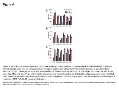
Figure 4 Figure 4. Replication of influenza viruses in vitro. MDCK cells (A), primary normal human
by jasmine
Bravo-Vasquez N, Karlsson EA, Jimenez-Bluhm P, Mel...
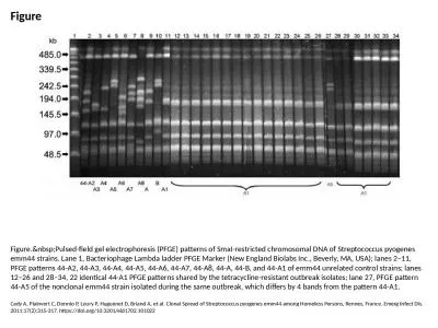
Figure Figure. Pulsed-field gel electrophoresis (PFGE) patterns of SmaI-restricted chromos
by white
Cady A, Plainvert C, Donnio P, Loury P, Huguenet D...
Figure 1 Figure 1. Selection of tuberculosis cases for analysis of sputum-culture conversion, Unite
by madeline
Scott C, Cavanaugh JS, Silk BJ, Ershova J, Mazurek...
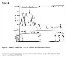
Figure 1 Figure 1. Fever and clinical course, 62-year-old woman.
by edolie
Mahara F. Japanese Spotted Fever: Report of 31 Cas...
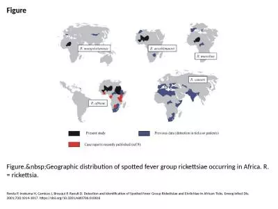
Figure Figure. Geographic distribution of spotted fever group rickettsiae occurring in Afr
by delilah
Parola P, Inokuma H, Camicas J, Brouqui P, Raoult ...
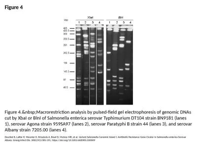
Figure 4 Figure 4. Macrorestriction analysis by pulsed-field gel electrophoresis of genomi
by amber
Doublet B, Lailler R, Meunier D, Brisabois A, Boyd...
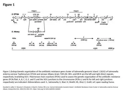
Figure 1 Figure 1. Genetic organization of the antibiotic resistance gene cluster of Salmo
by bella
Doublet B, Lailler R, Meunier D, Brisabois A, Boyd...
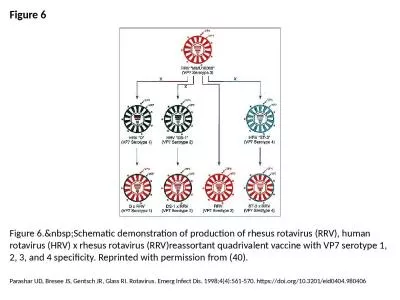
Figure 6 Figure 6. Schematic demonstration of production of rhesus rotavirus (RRV), human
by callie
Parashar UD, Bresee JS, Gentsch JR, Glass RI. Rota...
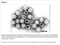
Figure 1 Figure 1. Rotavirus particles visualized by immune electron microscopy in stool f
by olivia
Parashar UD, Bresee JS, Gentsch JR, Glass RI. Rota...
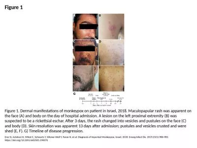
Figure 1 Figure 1. Dermal manifestations of monkeypox on patient in Israel, 2018. Maculopapular ras
by danya
Erez N, Achdout H, Milrot E, Schwartz Y, Wiener-We...
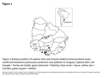
Figure 1 Figure 1. Locations of capture sites and enzyme-linked immunosorbent assay–conf
by natalie
Delfraro A, Clara M, Tomé L, Achaval F, Levis S, ...
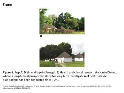
Figure Figure. A) Dielmo village in Senegal. B) Health and clinical research station in Di
by ariel
Parola P, Diatta G, Socolovschi C, Mediannikov O, ...
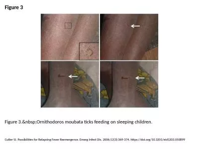
Figure 3 Figure 3. Ornithodoros moubata ticks feeding on sleeping children.
by tremblay
Cutler SJ. Possibilities for Relapsing Fever Reeme...
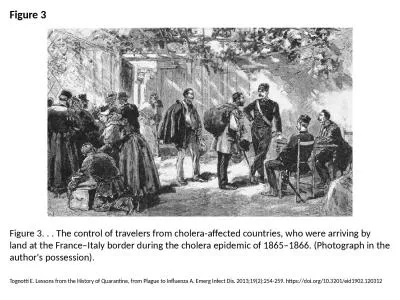
Figure 3 Figure 3. . . The control of travelers from cholera-affected countries, who were arriving
by rodriguez
Tognotti E. Lessons from the History of Quarantine...
Figure 3 Figure 3. Relative risks for employees at various machines on the factory floor i
by ava
van Woerden HC, Mason BW, Nehaul LK, Smith R, Salm...
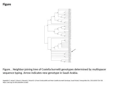
Figure Figure. . Neighbor-joining tree of Coxiella burnetii genotypes determined by multispacer seq
by blanko
Angelakis E, Johani S, Ahsan A, Memish Z, Raoult D...
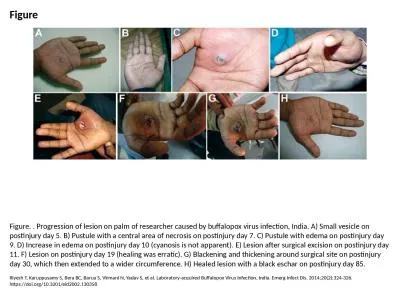
Figure Figure. . Progression of lesion on palm of researcher caused by buffalopox virus infection,
by eddey
Riyesh T, Karuppusamy S, Bera BC, Barua S, Virmani...
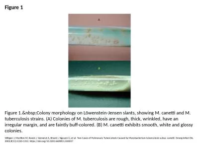
Figure 1 Figure 1. Colony morphology on Löwenstein-Jensen slants, showing M. canetti and
by ethlyn
Miltgen J, Morillon M, Koeck J, Varnerot A, Briant...
Load More...