Explore
Featured
Recent
Articles
Topics
Login
Upload
Featured
Recent
Articles
Topics
Login
Upload
Search Results for 'Figure-Cash'
Figure-Cash published presentations and documents on DocSlides.
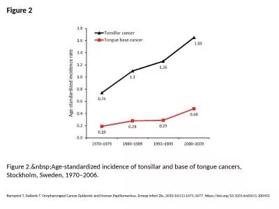
Figure 2 Figure 2. Age-standardized incidence of tonsillar and base of tongue cancers, Sto
by wang
Ramqvist T, Dalianis T. Oropharyngeal Cancer Epide...
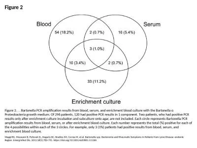
Figure 2 Figure 2. . . Bartonella PCR amplification results from blood, serum, and enrichment blood
by everly
Maggi RG, Mozayeni B, Pultorak EL, Hegarty BC, Bra...
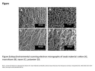
Figure Figure. Environmental scanning electron micrographs of swab material: cotton (A), m
by cecilia
Rose L, Jensen BJ, Peterson A, Banerjee SN, Arduin...
Figure 1 Figure 1. Verification of toxin gene deletions and the genetic structure of the construct
by elina
Plaut RD, Staab AB, Munson MA, Gebhardt JS, Klimko...
Figure 1 Figure 1. Lesions of the patient infected with Babesia divergens 1 day after hosp
by samantha
Haapasalo K, Suomalainen P, Sukura A, Siikamäki H...
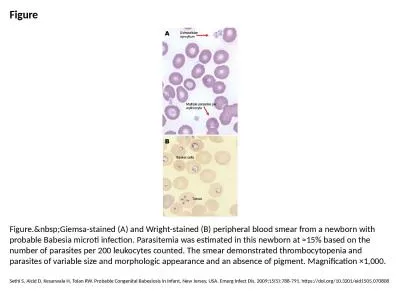
Figure Figure. Giemsa-stained (A) and Wright-stained (B) peripheral blood smear from a new
by deena
Sethi S, Alcid D, Kesarwala H, Tolan RW. Probable ...
Figure 2 Figure 2. A) Electron microscopy scan of peripheral blood. B) Transmission electr
by emma
O'Rourke LG, Pitulle C, Hegarty BC, Kraycirik S, K...
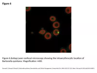
Figure 6 Figure 6. Laser confocal microscopy showing the intraerythrocytic location of Bar
by hadly
Foucault C, Brouqui P, Raoult D. Bartonella quinta...
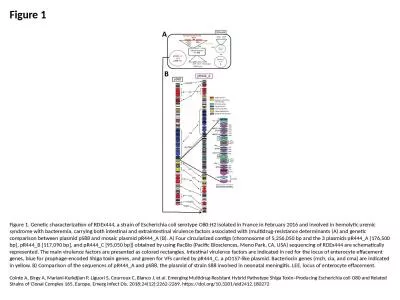
Figure 1 Figure 1. Genetic characterization of RDEx444, a strain of Escherichia coli serotype O80:H
by taylor
Cointe A, Birgy A, Mariani-Kurkdjian P, Liguori S,...
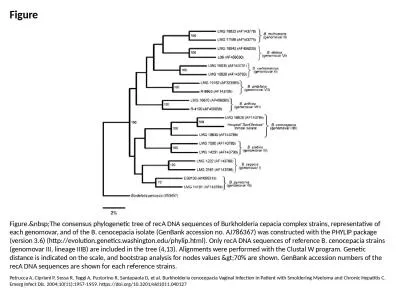
Figure Figure. The consensus phylogenetic tree of recA DNA sequences of Burkholderia cepac
by martin
Petrucca A, Cipriani P, Sessa R, Teggi A, Pustorin...
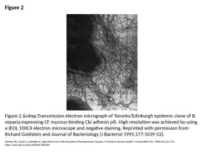
Figure 2 Figure 2. Transmission electron micrograph of Toronto/Edinburgh epidemic clone of
by danya
Holmes AH, Govan J, Goldstein R. Agricultural Use ...
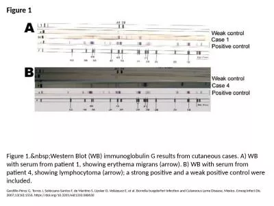
Figure 1 Figure 1. Western Blot (WB) immunoglobulin G results from cutaneous cases. A) WB
by ida
Gordillo-Pérez G, Torres J, Solórzano-Santos F, ...
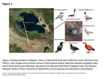
Figure 1 Figure 1. Location in Qinghai, China, of dead birds that were tested for avian in
by smith
Li Y, Liu L, Zhang Y, Duan Z, Tian G, Zeng X, et a...
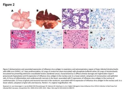
Figure 2 Figure 2. Lesions and associated expression of influenza virus antigen in respira
by joy
Reperant LA, van Amerongen G, van de Bildt MW, Rim...
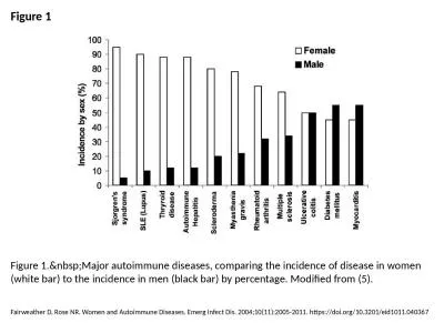
Figure 1 Figure 1. Major autoimmune diseases, comparing the incidence of disease in women
by bitsy
Fairweather D, Rose NR. Women and Autoimmune Disea...
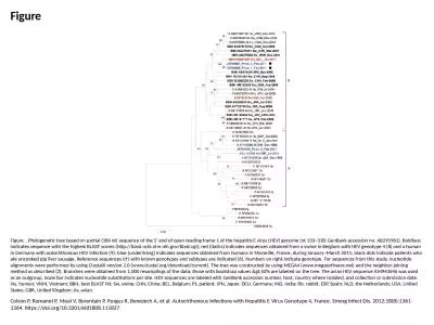
Figure Figure. . Phylogenetic tree based on partial (186 nt) sequence of the 5′ end of open readi
by eliza
Colson P, Romanet P, Moal V, Borentain P, Purgus R...
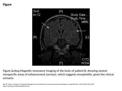
Figure Figure. Magnetic resonance imaging of the brain of patient B, showing several nonsp
by angelina
Kee AC, Yang S, Tambyah P. Atypical Chikungunya Vi...
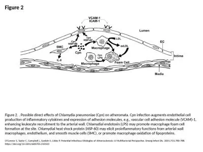
Figure 2 Figure 2. . Possible direct effects of Chlamydia pneumoniae (Cpn) on atheromata. Cpn infec
by cora
O'Connor S, Taylor C, Campbell L, Epstein S, Libby...
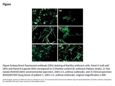
Figure Figure. Direct fluorescent-antibody (DFA) staining of Bacillus anthracis cells. Pan
by ruby
De BK, Bragg SL, Sanden GN, Wilson KE, Diem LA, Ma...
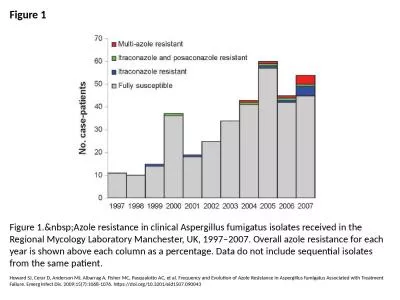
Figure 1 Figure 1. Azole resistance in clinical Aspergillus fumigatus isolates received in
by smith
Howard SJ, Cerar D, Anderson MJ, Albarrag A, Fishe...
Figure 4 Figure 4. Diagnostic algorithm incorporating the chest radiographic appearance and results
by daisy
Denning DW, Page ID, Chakaya J, Jabeen K, Jude CM,...
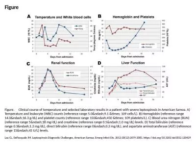
Figure Figure. Clinical course of temperature and selected laboratory results in a patient with se
by tremblay
Lau CL, DePasquale JM. Leptospirosis Diagnostic Ch...
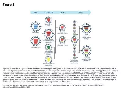
Figure 2 Figure 2. Illustration of original reassortment events of novel highly pathogenic avian in
by megan
El-Shesheny R, Barman S, Feeroz MM, Hasan M, Jones...
Figure 1 Figure 1. Global movement of wild birds (adapted from [8]) and geographic distribution of
by fanny
El-Shesheny R, Barman S, Feeroz MM, Hasan M, Jones...
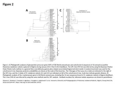
Figure 2 Figure 2. A) Phylogenetic analyses of glycoprotein precursor gene (GPC) of Old World arena
by molly
Olayemi A, Obadare A, Oyeyiola A, Igbokwe J, Fasog...
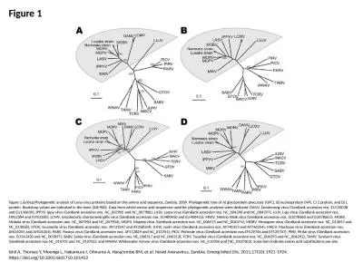
Figure 1 Figure 1. Phylogenetic analysis of Luna virus proteins based on the amino acid se
by delcy
Ishii A, Thomas Y, Moonga L, Nakamura I, Ohnuma A,...
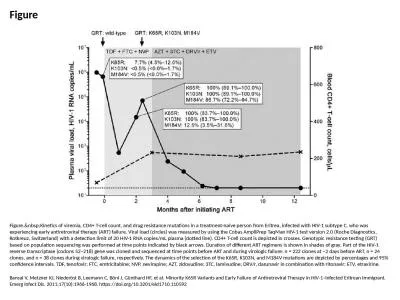
Figure Figure. Kinetics of viremia, CD4+ T-cell count, and drug resistance mutations in a
by amber
Bansal V, Metzner KJ, Niederöst B, Leemann C, Bö...
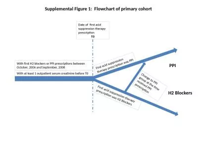
With at least 1 outpatient serum creatinine before T0
by elysha
With first H2 blockers or PPI prescriptions betwee...
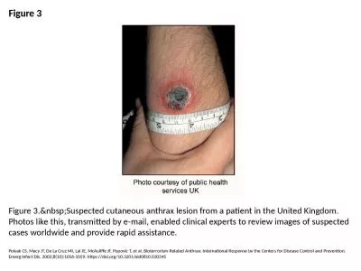
Figure 3 Figure 3. Suspected cutaneous anthrax lesion from a patient in the United Kingdom
by megan
Polyak CS, Macy JT, De La Cruz MI, Lai JE, McAulif...
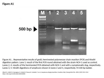
Figure A1 Figure A1. . Representative results of groEL heminested polymerase chain reaction (PCR) a
by williams
Alberti A, Addis M, Sparagano O, Zobba R, Chessa B...
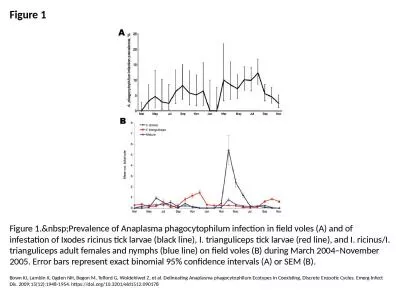
Figure 1 Figure 1. Prevalence of Anaplasma phagocytophilum infection in field voles (A) an
by ceila
Bown KJ, Lambin X, Ogden NH, Begon M, Telford G, W...
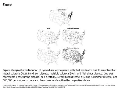
Figure Figure. Geographic distribution of Lyme disease compared with that for deaths due to amyotro
by sylvia
Forrester JD, Kugeler KJ, Perea AE, Pastula DM, Me...
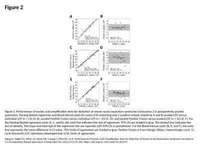
Figure 2 Figure 2. Performance of nucleic acid amplification tests for detection of severe acute re
by bitsy
Wang H, Hogan CA, Miller JA, Sahoo MK, Huang C, Mf...
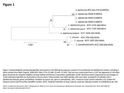
Figure 1 Figure 1. Neighbor-joining phylogenetic tree based on 16S rRNA gene sequence anal
by faith
Hall AJ, Cassiday PK, Bernard KA, Bolt F, Steigerw...
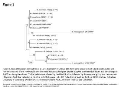
Figure 1 Figure 1. Neighbor-joining tree of a 1,341-bp region of unique 16S rRNA gene sequ
by lydia
Simmon KE, Brown-Elliott BA, Ridge PG, Durtschi JD...
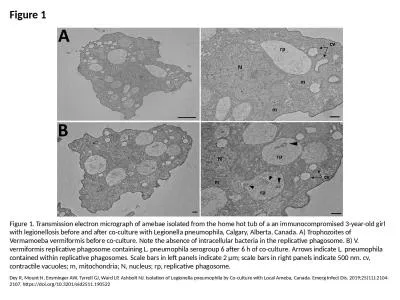
Figure 1 Figure 1. Transmission electron micrograph of amebae isolated from the home hot tub of a a
by joanne
Dey R, Mount H, Ensminger AW, Tyrrell GJ, Ward LP,...
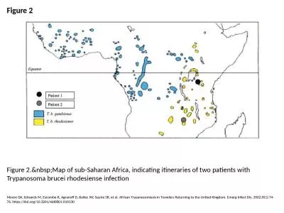
Figure 2 Figure 2. Map of sub-Saharan Africa, indicating itineraries of two patients with
by eve
Moore DA, Edwards M, Escombe R, Agranoff D, Bailey...
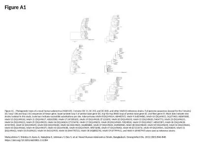
Figure A1 Figure A1. . Phylogenetic trees of a novel human adenovirus (HAdV-65), 3 strains (DC 11,
by kimberly
Matsushima Y, Shimizu H, Kano A, Nakajima E, Ishim...
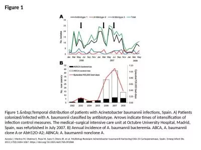
Figure 1 Figure 1. Temporal distribution of patients with Acinetobacter baumannii infectio
by taylor
Acosta J, Merino M, Viedma E, Poza M, Sanz F, Oter...
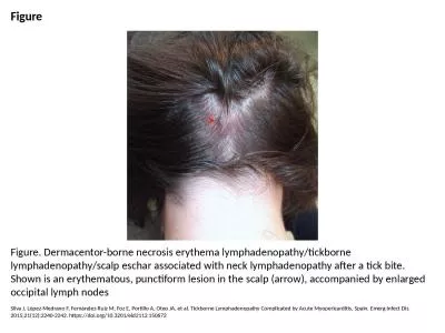
Figure Figure. Dermacentor-borne necrosis erythema lymphadenopathy/tickborne lymphadenopathy/scalp
by sophia
Silva J, López-Medrano F, Fernández-Ruiz M, Foz ...
Load More...


![Figure 1 Figure 1. Global movement of wild birds (adapted from [8]) and geographic distribution of](https://thumbs.docslides.com/1006702/figure-1-figure-1-global-movement-of-wild-birds-adapted-from-8-and-geographic-distribution-of.jpg)