Explore
Featured
Recent
Articles
Topics
Login
Upload
Featured
Recent
Articles
Topics
Login
Upload
Search Results for 'Micrograph'
Micrograph published presentations and documents on DocSlides.
MICROGRAPH ANALYSIS LAB
by min-jolicoeur
In lab book. Create 5 tables based on the followi...
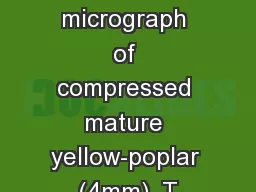
Figure C53. SEM micrograph of compressed mature yellow-poplar (4mm), T
by alida-meadow
Figure C54. SEM micrograph of compressed mature ye...
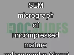
Figure C27. SEM micrograph of uncompressed mature yellow-poplar (4mm),
by alida-meadow
Figure C28. SEM micrograph of uncompressed mature ...
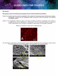
Micrographs Micrographs should be treated like photographs with the fo
by natalia-silvester
This information can be found on all micrographs t...
2015A Wet Mount Challenge
by celsa-spraggs
Instructions. Wet Mount PT: 2015A. Micrographs 1...
DDDs, Screening micrographs and CTF
by stefany-barnette
(Practical . work. ). . Vahid. . Abrishami. Int...
Figure C79. SEM micrograph of compressed mature southern pine, TRT1. .
by calandra-battersby
Figure C80. SEM micrograph of compressed mature so...
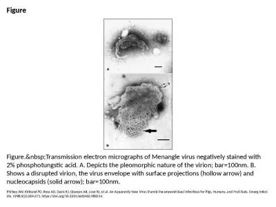
Figure Figure. Transmission electron micrographs of Menangle virus negatively stained with
by moises
Philbey AW, Kirkland PD, Ross AD, Davis RJ, Gleeso...
1 Design, Synthesis and Characterization
by bency
. of Glyc. o. l. i. pi. d. s. . and. Glycoclus. t...
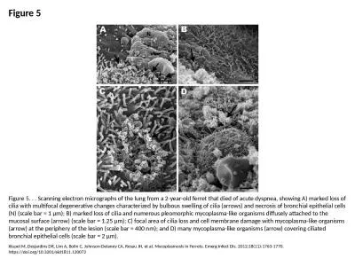
Figure 5 Figure 5. . . Scanning electron micrographs of the lung from a 2-year-old ferret that died
by taylor
Kiupel M, Desjardins DR, Lim A, Bolin C, Johnson-D...
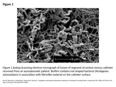
Figure 1 Figure 1. Scanning electron micrograph of lumen of segment of central venous cath
by lydia
Kim MJ, Bancroft E, Lehnkering E, Donlan RM, Masco...
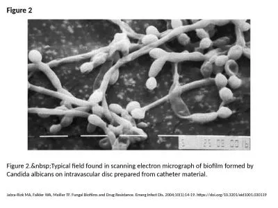
Figure 2 Figure 2. Typical field found in scanning electron micrograph of biofilm formed b
by melody
Jabra-Rizk MA, Falkler WA, Meiller TF. Fungal Biof...
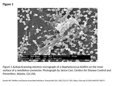
Figure 1 Figure 1. Scanning electron micrograph of a Staphylococcus biofilm on the inner s
by freya
Donlan RM. Biofilms and Device-Associated Infectio...
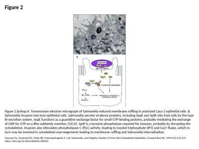
Figure 2 Figure 2. A. Transmission electron micrograph of Salmonella-induced membrane ruff
by victoria
Goosney DL, Knoechel DG, Finlay BB. Enteropathogen...
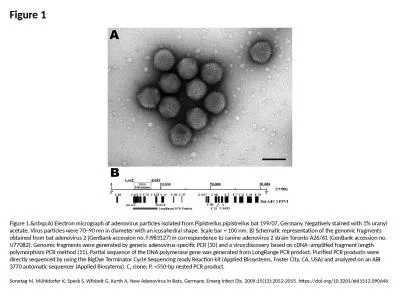
Figure 1 Figure 1. A) Electron micrograph of adenovirus particles isolated from Pipistrell
by lucy
Sonntag M, Mühldorfer K, Speck S, Wibbelt G, Kurt...
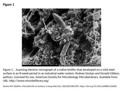
Figure 1 Figure 1. . Scanning electron micrograph of a native biofilm that developed on a mild stee
by victoria
Donlan RM. Biofilms: Microbial Life on Surfaces. E...
The Kidney Part Three – The Renal Corpuscle
by thomas
Digital Laboratory. It’s best to view this in . ...
Students Please spend a little time becoming familiar with PowerPoint operations. Running the Po
by oneill
1. To begin left click the slide show screen icon...
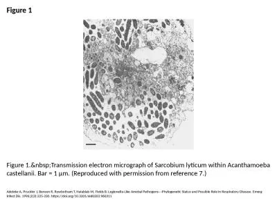
Figure 1 Figure 1. Transmission electron micrograph of Sarcobium lyticum within Acanthamoe
by elise
Adeleke A, Pruckler J, Benson R, Rowbotham T, Hala...
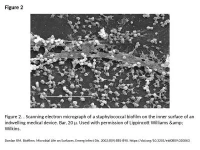
Figure 2 Figure 2. . Scanning electron micrograph of a staphylococcal biofilm on the inner surface
by patricia
Donlan RM. Biofilms: Microbial Life on Surfaces. E...
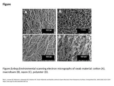
Figure Figure. Environmental scanning electron micrographs of swab material: cotton (A), m
by cecilia
Rose L, Jensen BJ, Peterson A, Banerjee SN, Arduin...
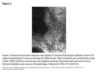
Figure 2 Figure 2. Transmission electron micrograph of Toronto/Edinburgh epidemic clone of
by danya
Holmes AH, Govan J, Goldstein R. Agricultural Use ...
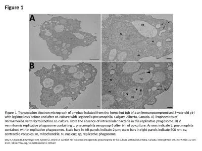
Figure 1 Figure 1. Transmission electron micrograph of amebae isolated from the home hot tub of a a
by joanne
Dey R, Mount H, Ensminger AW, Tyrrell GJ, Ward LP,...
Transmission Electron Micrographs of Negatively Stained Salmonella Typhimurium Flagella and Fimbria
by roxanne
Jejuni. Flagella. Authors: Rita Moyes, . Andra. ...
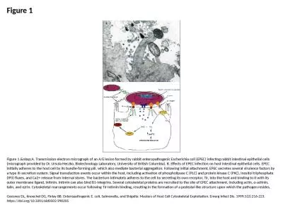
Figure 1 Figure 1. A. Transmission electron micrograph of an A/E lesion formed by rabbit e
by paige
Goosney DL, Knoechel DG, Finlay BB. Enteropathogen...
Lassa virus electron micrograph
by dorothy
Image courtesy, C.S. Goldsmith and M. Bowen (CDC)....
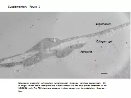
Supplementary figure 1 3d-astrocyte endothelial cell co-culture (uncompressed). Astrocytes constitu
by lucy
hCMEC. /D3 cells. The TEM shows one astrocyte in d...
Fellowshiptrained expertise inMohs micrographic surgery fortreatment
by lily
149Close coordination with Plastic Surgeryat the B...
Keywords - cell membrane; cell wall; nucleus; nucleolus; cytoplasm; mitochondria; ribosome; endoplasmic reticulum (smooth and rough); Golgi body; lysosome; vesicles
by myesha-ticknor
Keywords - cell membrane; cell wall; nucleus; nuc...
SEM & TEM in Polymer Characterization
by cheryl-pisano
Scanning Electron Microscopy (SEM). Uses. Sample ...
Preparation and Bioactivity evaluation of
by min-jolicoeur
bioresorbable. biphasic calcium phosphate . micr...
Ch 6: Tour of the Cell 2016
by phoebe-click
Chapter 6: Cells. From Topic 1.1. Nature of scien...
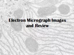
Electron Micrograph Images and Review
by sherrill-nordquist
Prokaryotic. Pili. Eukaryotic Animal Cell. Mitoch...
Topic 1.2 and 1.3 Review
by kittie-lecroy
1.2 Ultrastructure of cells. Essential Idea. : Eu...
1 .2 Ultrastructure of cells
by mitsue-stanley
Essential idea: Eukaryotes have a much more compl...
1. Explain how this animal’s cells will transform starch
by tatyana-admore
organelles. that are involved and the . processe...
1 .2 Ultrastructure of cells
by ellena-manuel
Essential idea: Eukaryotes have a much more compl...
Biology 1110
by celsa-spraggs
Principles of Biology. Biology 1110 Laboratory. L...
Preparation and Bioactivity evaluation of
by pamella-moone
bioresorbable. biphasic calcium phosphate . micr...
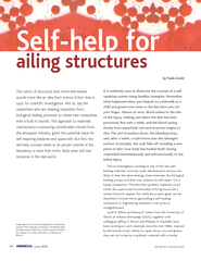
by Paula Gould Image above is an optical micrograph of
by olivia-moreira
Microcapsules containing a red healing agent are ...
Load More...