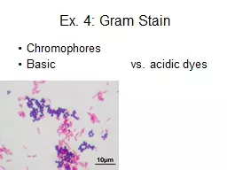
PPT-Ex. 4: Gram Stain Chromophores
Author : min-jolicoeur | Published Date : 2018-12-16
Basic vs acidic dyes Differential Stain Primary stain stains all cells on slide Decolorizing step removes stain from certain types of cells Counterstain stains the
Presentation Embed Code
Download Presentation
Download Presentation The PPT/PDF document "Ex. 4: Gram Stain Chromophores" is the property of its rightful owner. Permission is granted to download and print the materials on this website for personal, non-commercial use only, and to display it on your personal computer provided you do not modify the materials and that you retain all copyright notices contained in the materials. By downloading content from our website, you accept the terms of this agreement.
Ex. 4: Gram Stain Chromophores: Transcript
Basic vs acidic dyes Differential Stain Primary stain stains all cells on slide Decolorizing step removes stain from certain types of cells Counterstain stains the decolorized cells. Gram Stain Reaction. The Gram stain reaction, named after the Danish bacteriologist Hans Christian Gram, was originally devised in 1882.. Gram staining is based on the ability of the bacterial cell wall to retain the crystal violet dye. . Cheng . Zhang. Thurs 1 Dec 11. MM Tutorial. Gram staining. Fixed film (heat kill bacteria etc...). Methyl violet . Lugol’s. /gram’s iodine. Decolourise with acetone. Methyl red . . STAIN 2 (counter stain): pink-red colour. A stain is a substance that adheres to a cell, giving the cell color. .. The presence of color gives the cells significant contrast so are much more visible. . Different stains have different affinities for different organisms, or different parts of . Gram stain . Ziehl. . Neelsen. stain. . Requires the use of at least 3 chemical reagents.. Primary Stain. : . stain all the cells.. Decolorizing agent: . may or may not remove the primary stain from the entire the cell or only from certain cell structure. . Clinical Bacteriology II. CLS 413. Mrs. . Deemah. . Dabbagh. CLS Department. College of Applied Medical Sciences. King Saud University. Course Content. Enterobacteriacae. Lactose Fermenters:. Escherichia coli. BIOLOGY & BIOTECHNOLODY DEPARTMENT. In our laboratory, . bacterial morphology . (form and structure) may be examined in two ways:. By observing . living unstained organisms. (. wet mount. ).. 2. By . A stain is a substance that adheres to a cell, giving the cell color. .. The presence of color gives the cells significant contrast so are much more visible. . Different stains have different affinities for different organisms, or different parts of . Gram stain . Ziehl. . Neelsen. stain. . Requires the use of at least 3 chemical reagents.. Primary Stain. : . stain all the cells.. Decolorizing agent: . may or may not remove the primary stain from the entire the cell or only from certain cell structure. . Differentiate bacteria into two large groups (the Gram Positive and the Gram negative).. Gram status is important in medicine; the presence or absence of a cell wall will change the bacterium's susceptibility to some antibiotics. Differentiate bacteria into two large groups (the Gram Positive and the Gram negative).. Gram status is important in medicine; the presence or absence of a cell wall will change the bacterium's susceptibility to some antibiotics.. . By. Dr. . Shnyar. Hamid Qadir. Light Microscopy. Direct examination of stained or unstained preparations by . light (bright-field) microscopy. is particularly useful for detection of bacteria, fungi, and parasites.. Faculty: Dr. Rakesh Sharda OBJECTIVES OF STAINING Improves visibiltiy by greater contrast between the organism and the background, differentiate various morphological types (by shape, size, arrangeme Lab # 6. NEISSERIA SPECIES. Contains many species; 2 are human pathogens. Neisseria . gonorrhoea. (gonococcus). Neisseria . meningitidis. (. meningococcus. ). Other species are normal flora that inhabit the upper respiratory tract of humans.. Gram Positive. Gram Negative. Gram-Positive bacteria. Gram-Negative bacteria. Cell Wall. A single-layered, smooth cell wall. A double-layered, wavy cell-wall. Cell Wall thickness. The thickness of the cell wall is 20 to 80 .
Download Document
Here is the link to download the presentation.
"Ex. 4: Gram Stain Chromophores"The content belongs to its owner. You may download and print it for personal use, without modification, and keep all copyright notices. By downloading, you agree to these terms.
Related Documents