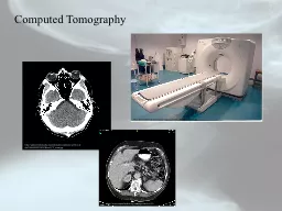
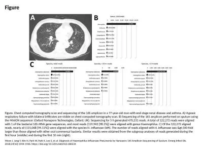
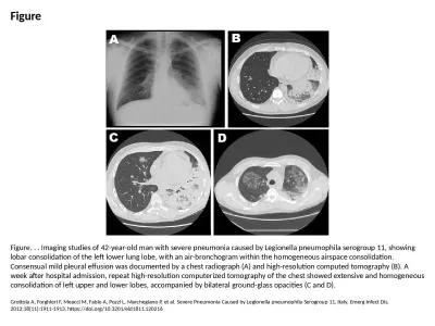
PPT-Figure Figure. . . Computed tomography scan showing a cavity (arrow) in the left lung

Author : kole295 | Published Date : 2024-09-18

Tortoli E Mariottini A Pierotti P Simonetti TM Rossolini G Mycobacterium yongonense in Pulmonary Disease Italy Emerg Infect Dis 2013191119021904 httpsdoiorg103201eid1911130911

Presentation Embed Code

Download Presentation

Download Presentation The PPT/PDF document "Figure Figure. . . Computed tomography s..." is the property of its rightful owner. Permission is granted to download and print the materials on this website for personal, non-commercial use only, and to display it on your personal computer provided you do not modify the materials and that you retain all copyright notices contained in the materials. By downloading content from our website, you accept the terms of this agreement.

Figure Figure. . . Computed tomography scan showing a cavity (arrow) in the left lung: Transcript


Tortoli E Mariottini A Pierotti P Simonetti TM Rossolini G Mycobacterium yongonense in Pulmonary Disease Italy Emerg Infect Dis 2013191119021904 httpsdoiorg103201eid1911130911. However perhaps we should broaden our horizons and look back through the centuries to obtain a more complete view of the development of human thought and aspirations which led to the invention of a device without which it would be dif64257cult to im Earls MD FSCCT GB John Mancini MD Koen Nieman MD PhD Gianluca Pontone MD Gilbert L Raff MD FSCCT CoChair University of British Columbia Vancouver Canada University of Texas Southwestern Medical Center Dallas Texas University of Erlangen Erlangen 20.. A teacher has had 2 weeks of back pain radiating down his posterior left leg. Which scan is the best for demonstrating a herniated disc?. MRA. CAT scan. PET scan. T1 MRI. T2 MRI. END OF QUIZ #3. Computed Tomography. Gerald R. Aben, MD FACR. Department of Radiology. College of Osteopathic Medicine. 2/13/2012. 1. Department of Radiology. Computed Tomography. Ionizing radiation. Requires concern and careful utilization. Limitations of Radiography. 3D body rendered in 2D. Structures superimposed on image. Structure of interest viewed through underlying / overlying structures. Multiple views used to adequately visualize structure.. http://upload.wikimedia.org/wikipedia/commons/archive/d/da/20060904231838!Head_CT_scan.jpg. http://. www.capitalhealth.org/subpage.cfm?ref. =36. Computed Tomography. . - Introduction. . Computed . Tomography. SR, JNMC, ALIGARH. Pleural cavity . The thoracic cavity is divided into three compartments . :. . right and left lateral compartments and . middle compartment. . . Each . lateral compartment is occupied by . Physics department. Calculation The Risk of Computed Tomography. Prepared by: Rayan F .Hassan. Fatima A .Mahmud. Supervised by:Msc.Rozhan. Outline. Objective. Introduction. Radiation dose from CT scans. Printe 09/01/2000 LAST UPDATED 05/17/2007 A Page 1of 2 9/16/2011 p Published:, August 2005 , June 2006 , July 2007 References: A merican College of Cardiology/American Heart Association Expert Consen 182[9-13].Recent studies have documented the abilityof this methodology to track progression or regres-sionof atherosclerosis using cholesterol-reducingmedications [14,15].Recent advances in spiral CT Website: www.certpot.com Certification Dump Prove Your Expertise in IT and Software Technologies Moon J, Jang Y, Kim N, Park W, Park K, Lee S, et al. Diagnosis of Haemophilus influenzae Pneumonia by Nanopore 16S Amplicon Sequencing of Sputum. Emerg Infect Dis. 2018;24(10):1944-1946. https://doi.org/10.3201/eid2410.180234. Grottola A, Forghieri F, Meacci M, Fabio A, Pozzi L, Marchegiano P, et al. Severe Pneumonia Caused by Legionella pneumophila Serogroup 11, Italy. Emerg Infect Dis. 2012;18(11):1911-1913. https://doi.org/10.3201/eid1811.120216. Pettit AC, Jahangir AA, Wright PW. Mycobacterium doricum Osteomyelitis and Soft Tissue Infection. Emerg Infect Dis. 2011;17(11):2075-2077. https://doi.org/10.3201/eid1711.110460.

Download Document

Here is the link to download the presentation.
"Figure Figure. . . Computed tomography scan showing a cavity (arrow) in the left lung"The content belongs to its owner. You may download and print it for personal use, without modification, and keep all copyright notices. By downloading, you agree to these terms.

Related Documents