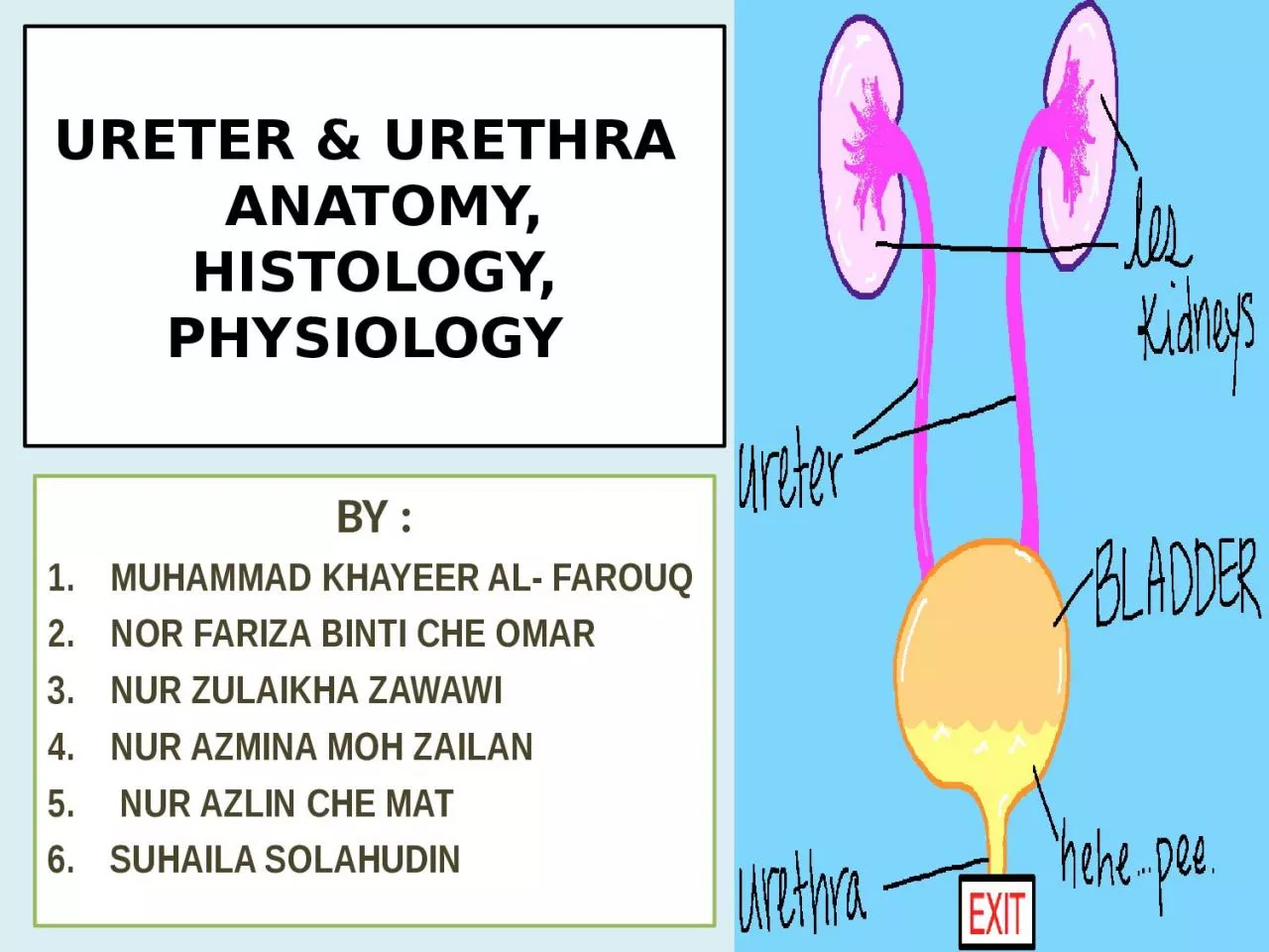
/
URETER & URETHRA   ANATOMY, HISTOLOGY, PHYSIOLOGY URETER & URETHRA   ANATOMY, HISTOLOGY, PHYSIOLOGY

URETER & URETHRA ANATOMY, HISTOLOGY, PHYSIOLOGY - PowerPoint Presentation

harper
harper . @harper
Follow
367 views
Uploaded On 2022-06-14

URETER & URETHRA ANATOMY, HISTOLOGY, PHYSIOLOGY - PPT Presentation

BY MUHAMMAD KHAYEER AL FAROUQ NOR FARIZA BINTI CHE OMAR NUR ZULAIKHA ZAWAWI NUR AZMINA MOH ZAILAN NUR AZLIN CHE MAT SUHAILA SOLAHUDIN ANATOMY OF THE URETER BY MUHAMMAD KHAYEER AL FAROUQ ID: 917902

ureter urethra epithelium urine urethra ureter urine epithelium male histology part female layer opening flow propria bladder lumen muscular

Share:

Link:

Embed:


Presentation Transcript

Slide1

URETER & URETHRA ANATOMY, HISTOLOGY, PHYSIOLOGY

BY :MUHAMMAD KHAYEER AL- FAROUQNOR FARIZA BINTI CHE OMARNUR ZULAIKHA ZAWAWINUR AZMINA MOH ZAILAN NUR AZLIN CHE MATSUHAILA SOLAHUDIN

Slide2

ANATOMY OF THE URETERBY : MUHAMMAD KHAYEER AL- FAROUQ D11B040

Slide3

The ureter is a muscular tube, which passes caudally In the retroperitoneal along the dorsal body wall. Reaching the pelvic cavity, it turns medially to enter the broad uterine ligament in the female, and yto the mesoductus deferens in the male.Ureter ends by inserting into the dorsolateral surface of the urinary bladder within the lateral ligament of the bladder.

Slide4

Urogenital system of cat Ventral aspect

URETER

Slide5

ANATOMY OF URETHRA BY : NOR FARIZA BINTI CHE OMARD11B041

Slide6

Slide7

Female urethra Female urethra extends caudally on the pelvic floor ventral to the reproductive tract. It passes obliquely through the wall of the vagina and opens with the external urethral opening (ostium urethrae externum) ventrally at the junction between vagina and vestibule.

Slide8

The length and diameter of the urethra varies considerably between the domestic mammals. Horse : short and wide and comparatively long in the dog were it opens on a small elevation flanked by two grooves.Cow and sow : urethralis muscle encloses the suburethral diverticulum which opens together with the urethra into the vagina.

Ureteral orifice Urethral m.External urethral orifices

Suburethral

diverticulum

Urethral crest

Slide9

Male urethra Extends from an internal opening at the bladder neck to an external opening at the end of the penis. It is divisible into a:Pelvis part Preprostatic portionProstatic portion Penile part

Slide10

Pelvic part Begins at the internal opening at the bladder neck. Preprostatic portion extends from the internal opening to the seminal hillock, an oval enlargement of the urethra crest which protrudes into the lumen of the urethra. Prostatic portionjoined by the deferent and vesicular ducts and passes through the prostate gland

Slide11

Penile part Begins at the ischial arch and is described with the penis

Slide12

Differentiate between male and female Female ShortWideElastic

MaleTortuousLong NarrowLess elastic

Slide13

Slide14

BY : NUR ZULAIKHA BINTI MAT ZAWAWID11A028HISTOLOGY OF URETER

Slide15

Histology Of UreterMagnification25x

100xKey1. Lumen 2. Transitional epithelium3. Mucous glands in lamina propria 4. Muscularis

Slide16

Microscopic structure of ureter

Slide17

Slide18

Histology of the Mucosa - inner layer of the ureter - consists of -epithelium - lamina propria.  Epithelium -The ureter

is lined by transitional epithelium. - The epithelial layer is avascular.  -does not contain lymphatics.  Lamina Propria - composed of areolar connective tissue.  - contains blood vessels and nerves.

Slide19

Histology of the Muscularis -The middle layer, the muscular coat  -consists of smooth muscle. -consist of - Circular muscular layer (smooth pink layer) - Longitudinal

muscular layer (bumpy purple layer).  -The main function of the muscularis is peristalsis which propels the urine. Adventitia/Serosa -The outer layer, the fibrous coat, is a supporting layer of fibrous connective tissue. -Adipose tissue is present in the adventitia. -A portion of the ureter has serosa covering it.

Slide20

URETER    Stained with haematoxylin and eosin 1 - tunica mucosa

2 - tunica submucosa 3 - tunica muscularis propria 4 - tunica adventitia 5 - transitional epithelium (urothelium) of the mucosa 6 - tunica propria of the mucosa URETERtransitional epithelium of the mucosa     Stained with haematoxylin and eosin 1 - transitional epithelium 2 - lamina propria of the mucosa

Slide21

HISTOLOGY OF URETHRABY : NUR AZMINA BT MOHD ZAILAND11A025

Slide22

Slide23

HISTOLOGY OF FEMALE URETHRA-The urethra in females is short. It is only about 1.5 inches.-The lining epithelium of the female urethra is pseudostratified

columnar epithelium and stratified squomous epithelium.

Slide24

HISTOLOGY OF FEMALE URETHRA

Slide25

HISTOLOGY OF MALE URETHRA-The urethra in the male is longer than the urethra of females. -The male urethra is 7 to 8 inches. -The male urethra functions as a conduit for urine and semen.

-The male urethra can be divided into 3 sections : prostatic urethra,membranous urethra, and spongy urethra.

Slide26

HISTOLOGY OF MALE URETHRA

Male urethra is lined by stratified columnar epithelium.

Slide27

HISTOLOGY OF PROSTATIC URETHRA (MALE)Stained with haematoxylin and eosin 1- transitional epithelium 2- tunica propria

of the mucosa of prostatic part of the urethra

Slide28

HISTOLOGY OF MEMBRANOUS URETHRA( MALE)The second part of the male urethra.This is a short segment.The lining epithelium of the membranous urethra is pseudostratified columnar epithelium.

Slide29

HISTOLOGY OF SPONGY URETHRAThe third part of the urethra.Spongy urethra is also called the penile urethra.The longest section of the male urethra.The lining epithelium of the spongy urethra is pseudostratified columnr epithelium which then transitions to stratified

squamous epithelium distally.

Slide30

PHYSIOLOGY OF URETERBY : NURAZLIN BINTI CHE MATD11A029

Slide31

Flow Of Urine In UreterMyogenic = Controlled by the longitudinal and circular muscle of ureter wallPeristaltic movement = waves of active muscular contraction, that move at near-constant speed along the ureter towards the bladder . This causes urine to flow from the ureter lumen to the urinary bladderUnaffected by drugs that block the sympathetic and parasympathetic nervous system .muscular wall constricts & becomes thinner during luminal enlargement ( urine present )

Slide32

Very low volume of urine at certain part of ureter or no urine at all

High volume of urine at certain part of ureterVolume of urine at certain part of ureter increases

Slide33

Diagram showing the different stages of ureter opening during urine flow in it

Slide34

1a, 2a, 3a, 4a

Pictures of cross section of ureter showing stages of ureter opening upon urine flow in it( light micrograph)1b,2b,3b,4bPhotograph of cross section of ureter showing various stages of ureter opening upon urine flow in it

Slide35

No urine present at certain part of ureter=At the end of a peristaltic movement or at rest between movements = The lumen is collapsed with its epithelial surfaces in contact with each other . The

lumen has a stellate form

Slide36

There are junctional complexes, comprising a zonula occludens, zonula adherens , and macula adhaerens or desmosome, between the lateral borders of the squamous cells . It is suggested that

this complex is t h e major obstacle to the free flow of water from the extracellular spaces into the hypertonic urine ( in lumen of ureter )

Slide37

Slide38

PHYSIOLOGY URETHRA BY : SUHAILA BINTI SOLAHUDIN D11A035

Slide39

Function of urethra both in male and female :Transport urine from the urinary bladder to the exterior of the bodySpecial function in male :Carries semen from the ejaculatory duct outwards through the penis.

Slide40

How the urine flow out of the body?When the sphincter muscle is relaxed. Urethral sphincter muscle is at the base of the bladderIt controls the release of urine from the

bladder into the urethraThen flows out of the meatus (opening at the end of the urethra) when urinating. 

Slide41

THANK YOU (^^,)

BLUE GLAUCUS