/
GAIT: NORMAL, ABNORMAL & ASSESSMENT GAIT: NORMAL, ABNORMAL & ASSESSMENT

GAIT: NORMAL, ABNORMAL & ASSESSMENT - PowerPoint Presentation

evans
evans . @evans
Follow
502 views
Uploaded On 2022-06-28

GAIT: NORMAL, ABNORMAL & ASSESSMENT - PPT Presentation

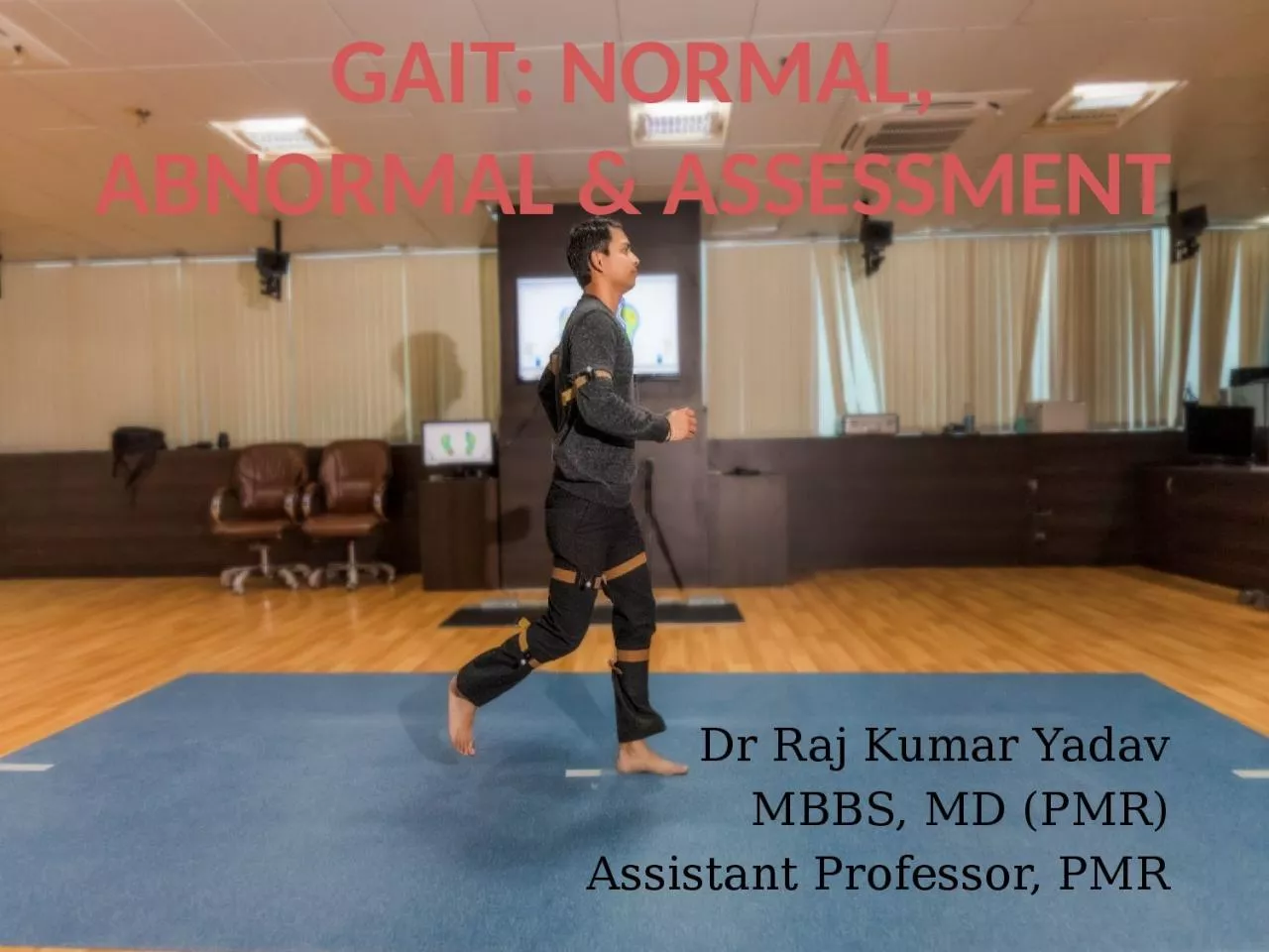
Dr Raj Kumar Yadav MBBS MD PMR Assistant Professor PMR GAIT 1 Normal Walking 2 Gait cycle phases temporal parameters 3 Determinants of gait 4 Kinematic amp kinetic analysis ID: 926519

phase gait knee swing gait phase swing knee stance limb hip flexion walking side foot contact ground length speed

Share:

Link:

Embed:


Presentation Transcript

Slide1

GAIT: NORMAL, ABNORMAL & ASSESSMENT

Dr Raj Kumar Yadav

MBBS, MD (PMR)

Assistant Professor, PMR

Slide2

GAIT

1- Normal Walking2- Gait cycle – phases, temporal parameters3- Determinants of gait4

- Kinematic & kinetic analysis

5

- Gait in young, elderly & women

6

-

Some abnormal gaits

7- Assessment – visual, video recording

8- Clinical Gait laboratory

Slide3

Walking

Walking

- complex interaction of different parts of body

- it’s advancement in the desired line of progression.

M

uscle act

-

this motion and forces are controlled

Normal walking –

-

weight

bearing stability and

progression

over the supporting

foot

optimal conservation

of physiologic energy.

Slide4

GAIT CYCLE :- Activity that occurs between heel strike of one extremity and subsequent heel strike same

side.

STANCE

PHASE :- Phase in which limb is in contact with the ground. (60

%)

SWING

PHASE :- Phase in which the foot is in air for limb advancement. (40

%)

DOUBLE SUPPORT: When

two extremities are in contact with the ground

simultaneously

- cadence (speed of walking) -

double support

- Absence

of double support -

running

Slide5

Slide6

DEFINITIONS

Initial contact: (0%) Instant the foot contacts the ground.

Loading response: (0-11%)

immediately

following initial contact -

lift of C/L extremity from

ground

weight

shift occurs

.

Slide7

Subphases of Stance phase

Mid- stance: (11-30%)

- lift

of C/L extremity from ground

- ankles

of both extremities are aligned in the frontal (coronal) plane

.

Terminal stance: (30-50%)

- ankle alignment in frontal plane - just prior to initial contact of C/L extremity.

Preswing

: (50-60%)

- initial contact of C/L extremity - prior lift of

Ipsilateral

extremity from ground.

Slide8

Sub phases of Swing phase

Initial swing: (60-73%) Lift of the extremity from ground - position of maximum knee flexion.Mid swing: (73-87%) Immediately following knee flexion - vertical tibia position.

Terminal swing: (87-100%) Following vertical tibia position

-

just prior to Initial contact.

Slide9

Slide10

Temporal Gait Parameters

Stride length: Linear distance between corresponding successive points of contact of the same foot

Highly variable - normalized by dividing it by leg length or total body height

increases as the speed increases.

Step length: opposite foot

- gait symmetry.

Slide11

Cadence: No. of steps/minuteVelocity (meters/minute): Distance covered in given time in the given direction.

Step width (width of walking base): Distance between the midpoints of the heel of two feetincreases - increased demand for side to side stability.

Slide12

Degree of toe-out:

Represents the angle of foot placementAngle

between the line of progression and the line intersecting the

centre

of heel and the second

toe

decreases

as the speed increases

Slide13

Temporal gait parameter

Average value

Velocity (m/s)

0.9 – 1.5

Cadence (steps/min)

90 - 135

Stride length (m)

1 – 1.5

Step length (cm)

38

Walking base (cm)

6 - 10

Degree of toe-out

Stance phase

60%

Swing phase

40%

Double limb support

20%

Slide14

Determinants of Gait (Saunders 1953)

Optimizations to minimize excursion of centre of gravity (COG), hence reduction of energy consumption

Pelvic rotation

Pelvic tilt

Knee flexion in stance

Ankle PF

Foot supination

Lateral displacement of the pelvis

Determinants 1 - 5 reduce displacement on the vertical plane (50%)

determinant 6 - horizontal plane (40%).

Slide15

GROUND REACTION FORCE (GRF)- When a person takes a step, forces are applied to the ground by the foot and by the ground to the foot (GRF)

equal but opposite GRFVector = sum of the force components in each direction (vertical, anteroposterior

and

mediolateral

axes)

typical pattern from initial contact to toe-off.

MOMENTS (Torque/ turning force)-

External forces - GRF, gravity and inertia - external moments about the joints.

Internal moments - moments generated by the muscles, joint capsules, and ligaments -

countract

the external forces

Slide16

Muscle activity

Slide17

Kinetics and Kinematics

Kinetics : Study of forces, moments, masses and accelerations, but without any detailed knowledge of the position or orientation of objects involved.

Kinematics : Describes motion, but without reference to forces involved.

Slide18

Trunk and Shoulder

Trunk along with shoulder girdle twists in opposite direction of pelvic twistTotal excursion of trunk is 7° and pelvic girdle 12°.

Total ROM of shoulder is 30

°

(24

°

of extension and 6

°

of flexion)

Center of gravity (COG) is located 5 cm anterior to second sacral vertebra

It is displaced 5 cm horizontally and 5 cm vertically during a gait cycle.

Slide19

Gait in children

Children have no heel strike, initial contact being made by flatfoot (2 yr)Very little stance phase knee flexion (2 yr)

Whole leg is externally rotated during swing phase (2 yr)

Walking base is wider (4 yr)

Absence of reciprocal arm swing (4 yr)

Stride length and velocity are lower and cadence higher (15 yr)

Slide20

GAIT IN ELDERLY

Decreased stride length and cadenceIncrease in walking base

Reduction in total range of flexion and extension

of joints

GAIT IN WOMEN

Gait speed is slower

Step length is smaller

Increased cadence

Slide21

ABNORMAL GAIT

Any deviation from normal pattern of walkingCaused

motor system

skeletal

supports

neural

control

combination

of the

above.

Slide22

PAINFUL/ANTALGIC GAIT

HIGH STEPPAGE

GAIT

Avoidance of weight bearing on the affected limb

shortening of stance phase in that limb

weakness of ankle

dorsiflexors

excessive knee and hip flexion with toes pointing downwards in the swing phase

Slide23

VAULTING

Seen in limb length discrepancy, hamstring weakness or extension contractures of the kneeThe knee is hyper-extended and locked at end of stance phase and entire swing phase.So to clear the leg the patient goes up on the toes of the other leg to clear the affected limb.

Slide24

TRENDELENBURG GAIT

The gluteus medius during the stance phase, pulls the stance side pelvis over the supporting limb to prevent excessive pelvic drop in the opposite swing limb.

If the hip abductors are weakened, the opposite limb pelvis may drop excessively during swing phase.

To avoid this, the entire trunk shifts to the stance side to bring the stance pelvis on to the supporting limb.

This is known as gluteus

medius

lurch or

trendelenburg

gait.

Slide25

MYOPATHIC GAIT

If both hip abductors are weak, the trunk sways from side to side during the stance phase to bring the pelvis level on the supporting limb.waddling gait.

muscular dystrophies

accompanied by excess lumbar

lordosis

to compensate for hip extensor weakness.

Slide26

HEMIPLEGIC GAIT

In extensor synergy -heel strike is missing and patient lands on forefoot

Since hip and knee are kept extended throughout the gait cycle, there is relative limb lengthening and hence

circumduction

or hip hiking is used for clearance

Toe drag may be present in swing phase

Swing phase is longer on the affected limb

Decreased arm swing on the affected side.

If flaccid paralysis or flexor synergy is present

knee buckling and instability

Slide27

FESTINATING/PROPULSIVE GAIT

Lack of arm swingShort, quick steps with increasing speed Cannot stop abruptly or change directionsStooped posture

Seen in

Parkinsonism

Carbon monoxide poisoning

Slide28

ATAXIC GAIT

Seen in cerebellar lesionsDysmetria and inco

-ordination

Staggering and lack of smooth movements (reeling or drunken gait)

Falls to the side of lesion

Compensated by wide-based gait to increase base of stability

Slide29

STOMPING GAIT

Seen in sensory ataxia Gait with heavy heel strikes, forceful knee extension and improper foot placement as well as a postural instabilityUsually worsened when the lack of proprioceptive input cannot be compensated for by visual input, such as in poorly lit environments.

Friedreich’s

ataxia, pernicious

anemia

,

tabes

dorsalis

, spinal cord pathologies

Slide30

CEREBRAL PALSY GAIT

Crouch gaitHip and knee increased flexion throughout stance with ankle

dorsiflexion

Due to hamstring tightness

Jump

knee gait

Flexion at hip and knee and ankle

equinus

is characteristic of this gait

Slide31

GAIT IN CEREBRAL PALSY

Stiff knee gait excess knee extension throughout swing

Has to use

circumduction

or vaulting

Due to increased rectus

femoris

activity in swing phase

Recurvatum

knee

Due to triceps spasticity or hamstrings transfer

Leads to increased knee extension in mid & late stance

Slide32

SCISSORING GAIT

Spasticity of the hip adductors with relative weakness of hip abductors and secondary changes in the hip gives rise torigidity and excessive adduction of the leg in swing

plantar flexion of the ankle

increased flexion at the knee

adduction and internal rotation at the hip

Diplegic

CP, Spinal cord pathologies

Slide33

METHODS OF GAIT ANALYSIS

Slide34

VISUAL GAIT ANALYSIS

The simplest form of gait analysis.Look for: Symmetry and smoothness of movements

Balance

Degree

of effort

Motion of specific segments

Gait parameters

Gait should be observed from at least 3 angles (side, front & back)

Slide35

Limitations-

gives no permanent record

eyes

cannot observe high-speed

events

only

possible to observe movements not

forces

depends

entirely on the skill of the individual observer

.

Gait analysis walkwayLength – 10-12 m

Width

- visual - 3

m video recording - 4 m kinematic system - at least 6 m.

Slide36

ANALYSIS BY VIDEO RECORDING

Advantages-gives permanent recordcan observe high speed events

reduces the number of walks a subject needs to do

makes it possible to show the subject exactly how they are walking

makes it easier to teach visual gait analysis to someone else.

The majority of today’s domestic cameras are perfectly suitable for use in gait analysis

Slide37

Clinical Gait laboratory

A fully equipped clinical gait laboratory can be expected to posses a combined kinetic/kinematic systems, with ambulatory EMG, as well as facilities for making videotapes.

Equipment may also be available for measuring oxygen uptake or pressure beneath the feet

Slide38

KINEMATICS –

- Camera by using infrared radiations measures the position of the markers

FORCE

PLATFORM /

FORCEPLATE

- Usual

methods of displaying

force platform

data

is

the

butterfly diagram

Slide39

ELECTROMYOGRAPHY (EMG)

EMG measures the electrical activity of a contracting muscle during different phases of gait cycle 1- Surface electrodes- Not suitable for deep muscles like

iliopsoas

.

2-

Fine wire

electrodes-

3-

Needle electrodes-

Slide40

MEASURING ENERGY CONSUMPTION

Oxygen consumption- measurements of oxygen

uptake

while

not particularly pleasant for the subject (who has to wear face mask or mouth

piece)

Practical

Whole body

calorimetry

-

most accurate way

but quite impractical subject is kept in an insulated chamber for measuring the heat output of the bodyPhysiological Cost Index: less accurate

PCI =

(Walking HR – Resting HR)

Walking Speed in m/min

Slide41

Slide42

Slide43

Slide44

Slide45

Thank You.