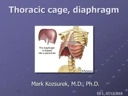
PPT-Dr. Rehan Clinical anatomy of thoracic cage and cavity-1

Author : eliza | Published Date : 2022-04-07

At the end of this session the student should be able to Discuss briefly anatomical changes in thorax with ageing Describe needle and tube thoracostomy Identify

Presentation Embed Code

Download Presentation

Download Presentation The PPT/PDF document "Dr. Rehan Clinical anatomy of thoracic c..." is the property of its rightful owner. Permission is granted to download and print the materials on this website for personal, non-commercial use only, and to display it on your personal computer provided you do not modify the materials and that you retain all copyright notices contained in the materials. By downloading content from our website, you accept the terms of this agreement.

Dr. Rehan Clinical anatomy of thoracic cage and cavity-1: Transcript


At the end of this session the student should be able to Discuss briefly anatomical changes in thorax with ageing Describe needle and tube thoracostomy Identify indication of thoracotomy and structures encountered in performing it . 27 Copyright The McGrawHill Companies Inc Permission required for reproduction or display Sternoclavicular joint Acromioclavicular joint Clavicle Scapula Suprasternal notch Clavicular notch Manubrium Angle Body Xiphoid process Costal cartilages Cost Kristin Washburn. May 23, 2014. Conflicts of Interest. None. Outline. Background/history . Physiologic benefits . Current practice. Current Research . Fluoroscopy and . epidurograms. RCT. Timeline. 1885: Coring documented epidural anesthesia in animals. The first ribarticulates with the manubrium via a long piece of flexible cartilage, and may be palpated near the manubrium for about two centimeters below the inferior margin of the clavicle, and at But why does it work?. The Cage is named after Faraday, but why it works begins with Coulomb. Coulomb determined: Electric field E at radius r from stationary point charge Q is equal to…. ϵ. 0. is the permittivity of free space and . lumbar vertebrae . (5) bear greater loads, they are much stouter than their more superior cousins (the cervical and thoracic vertebrae).. The Vertebral Column. The . sacrum. is a single triangular body formed from a fusion of 5 separate vertebrae in-utero (during fetal development).. LISA PIROPATO, PT, DPT, ATC. NORTHEAST INDIANA SPORTS MEDICINE SYMPOSIUM. MARCH 25, 2017 . Learning objectives. Explain . the etiologies of thoracic outlet syndrome. . Differentiate . between . the types of vascular and neurologic thoracic outlet syndrome. . Department of Anatomy. Faculty of Medicine. Objectives. Gross structure, function & dysfunction. 1. . Describe . the component parts . of the diaphragm and state its functions . 2. Describe the . Part 2. Body Regions and Body Cavities. Body Regions. HEAD. : Face. Cranium. NECK. :. Cervical [anterior and posterior]. TRUNK. : Thorax. Abdomen. Pelvis. Back- Vertebral. Lumbar/Sacral. UPPER APPENDAGE. Sept. 8, 2015. Requirements / Assumptions. Provide . the. nominal drift field of 500V/cm . Withstand -180kV near the cathode. Define the drift distance between the APAs and CPAs to <1cm*. Use materials with comparable CTEs to that of stainless steel along the length of the TPC, minimal CTE in the drift direction. Text: Genesis 4. The rage cage #1. A ‘. Just. . Enough. ’ Dedication to God (Which Always Results in . Envy. ) . (vv1-5). The rage cage #1. Gen. 4:1. Now Adam knew Eve his wife, and she conceived and bore Cain, saying, “I have gotten a man with the help of the Lord.”. Mark Kozsurek, M.D., Ph.D.. ED I. ., 07/12/2018. Questions. . to. be . answered. :. How. . are. . the. . bones. of . the. . thorax. . connected. . together. ?. . → . the. . joints. of . SR, JNMC, ALIGARH. Pleural cavity . The thoracic cavity is divided into three compartments . :. . right and left lateral compartments and . middle compartment. . . Each . lateral compartment is occupied by . BY:. DR. Stella Selena. M.B.B.S., DGO.. Academy Director. Mother Teresa Institute Of. Pharmaceutical Education and Research. Two Branches Of Science. Anatomy. : Is the science of the body structures and their relationships among them. Skull, Vertebral Column, Hyoid Bone & Thoracic Cage. Functions. T. he Skeleton:. Supports. . – rigid framework to support soft tissue that weighs 5x the amount of the skeleton. Protects. . - Organs are protected by ribs cage & pelvis & marrow is protected; red .

Download Document

Here is the link to download the presentation.
"Dr. Rehan Clinical anatomy of thoracic cage and cavity-1"The content belongs to its owner. You may download and print it for personal use, without modification, and keep all copyright notices. By downloading, you agree to these terms.

Related Documents