
PSK4U related to the heart Cardiovascular System Composed of Heart Blood vessels Blood Cardiovascular System Functions Delivery of O 2 fuel and nutrients to the tissues of the body ID: 774937
Download Presentation The PPT/PDF document " The Cardiovascular System" is the property of its rightful owner. Permission is granted to download and print the materials on this web site for personal, non-commercial use only, and to display it on your personal computer provided you do not modify the materials and that you retain all copyright notices contained in the materials. By downloading content from our website, you accept the terms of this agreement.


Slide1
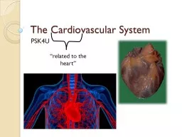
The Cardiovascular System
PSK4U
“related to the heart”
Slide2Cardiovascular System
Composed of:HeartBlood vesselsBlood
Slide3Cardiovascular System
Functions:Delivery of O2, fuel, and nutrients to the tissues of the bodyRemoval of CO2 and waste products from the tissues
Slide4Cardiovascular System
Functions:Maintenance of a constant body temperature (thermoregulation)Prevention of infection (immune function)
Slide5The Heart
Formed from myocardium, a specialized muscle tissueSurrounded by pericardium (tough protective sac); allows heart to expand and contract
Slide6The Heart
Epicardium lines outside of heart; endocardium lines inside of heart
Slide7The Heart
Made up of four separate chambers: atria (upper chambers) and ventricles (lower chambers)
Slide8The Heart
Ventricles are separated from atria by specialized valves that allow blood to flow only from atria into ventriclesCalled Atrioventricular (AV) valvesRight side-Tricuspid valve-3 flapsLeft side-Bicuspid (Mitral) valve-2 flaps Attached to papillary muscles by chordae tendinaePrevent inversion
Slide9The Heart
Semilunar valves-blood leaves the ventriclesPulmonary Semilunar ValveRight side of heartPrevents blood from flowing back from the pulmonary arteries into the right ventricleAortic Semilunar ValveLeft side of the heartSeparates the aorta from the left ventricle
Slide10The Heart
Considered a “double-pump” and is divided into the right and left heart; separated by the
interventricular
septum
Right heart:
pumps deoxygenated blood to the lungs (
pulmonary circulation
)
Very dark red, depicted as blue
Left heart:
Pumps oxygenated blood to the rest of the body (
systemic circulation
)
Bright red
Slide11Structures of the Heart
Common Structures
Structure of right side
Structure of left side
Chordae
tendinae
Superior and inferior vena cava
Aorta and thoracic (descending aorta)
Papillary muscles
Right atrium
Left atrium
Interventricular
septum
Right ventricle
Left ventricle
Pulmonary artery
Pulmonary vein
Tricuspid valve
Bicuspid (mitral) valve
Pulmonary valve
Aortic valve
Slide12The Internal Anatomy of the Heart
Aorta
Superior vena cava
Right pulmonary artery
Aortic semilunar valve
Right pulmonary veins
Right atrium
Pulmonary semilunar valve
Tricuspid valve
Right ventricle
Inferior vena cava
Left pulmonary artery
Left pulmonary veins
Left atrium
Bicuspid (mitral) valve
Left ventricle
Chordae
tendinae
Papillary muscles
Interventricular septum
Chordae
tendinae
Papillary muscles
Thoracic aorta (descending)
Slide13Path of Blood Through the Heart
Aorta
Superior vena cava
Right pulmonary artery
Aortic semilunar valve
Right pulmonary veins
Right atrium
Pulmonary semilunar valve
Tricuspid valve
Right ventricle
Inferior vena cava
Left pulmonary artery
Left pulmonary veins
Left atrium
Bicuspid (mitral) valve
Left ventricle
Chordae tendinae
Papillary muscles
Interventricular septum
Chordae tendinae
Papillary muscles
Thoracic aorta (descending)
Slide14Try These activities
Heart Quiz
Blood Flow
Quiz
This one is a bit harder
Slide15Systemic vs Pulmonary Circulation
Arteries carry blood away from the heart
Systemic circulation
(vast majority of body’s blood vessels)
Carry oxygenated blood from heart to the tissues
Pulmonary circulation
Carry deoxygenated blood from heart to lungs
Veins carry blood toward the heart
Systemic circulation
Carry deoxygenated blood from tissues back to heart
Pulmonary circulation
Carry oxygenated blood from lungs back to heart
Slide16Cardiac Muscle
Similar in structure to skeletal muscle
Interconnected and excitable
Allow passage of electrical signals
Allows the myocardium to contract as a unit
When a single cell is stimulated to contract it causes all other cardiac muscles to contract
SYNCYTIUM
Contraction of the heart leads to pumping of blood
Slide17The Heart – Electrical Conduction System
Sinoatrial (SA) node
Internodal
pathways
Bundle of His (AV bundle)
Atrioventricular (AV) node
Right and left bundle branches
Purkinje
fibres
Slide18Excitation of the Heart
Sinoatrial
node (SA node)
:
Specialized region of tissue found in wall of right atrium
Location where electrical signals are initiated (“pacemaker”)
Sets the basic rate of contraction
Modulated by the autonomic nervous system
Electrical signals are spread through both atria by
internodal
pathways
(top to bottom)
Slide19Excitation of the Heart
Atrioventricular
node (AV node)
:
Passes electrical signal from atria into
ventricles
Passes electrical signal to the bundle of His (
atrioventricular
bundle
)
Bundle of His pass electrical signal to the
Purkinje
fibres
Purkinje
fibres
pass electrical signal to the
myocardium
The myocardium of ventricles contract
(bottom to top)
Leads to contraction of the heart
Leads to the pumping of blood
Slide20The Electrical Activity of the Heart
Measured using an electrocardiogram (ECG)Graphical representation of electrical sequence of events occurring with each contraction of the heartEach wave generated during contraction is named:P wave: represents depolarization through the atriaSpreading of the electrical signal to contract through the atriaAtria is immediately repolarized –not visible in ECGQRS complex: represents depolarization of the ventricleT wave: represents repolarization of the ventricle
Slide21Slide22Coronary Circulation
Heart requires constant supply of O
2
, fuel and nutrients
Myocardial infarction
Blood supply to a region of the myocardium is cut off for a prolonged period of time or blood flow is reduced
Myocardium will die or become damaged
HEART ATTACK
Slide23Cardiac Cycle
Cardiac cycle
: series of events occurring through one heartbeat
Dramatic changes in pressure in the heart-measure in arteriesInvolves two phases:
Systole phase (contraction) Heart contracts and ejects blood
Diastole phase (relaxation)
Heart fills with blood
Slide24Summary of the Vascular System
Large veins
Medium veins
Venules
Large arteries
Medium arteries
Capillaries
Arteriole
Precapillary sphincters
Capillary bed
Slide25The Vascular System and Blood
Vascular System:A network of vessels that transport blood throughout the bodyEndothelium lines the inside of all vesselsVessels divided into four main categories:Arteriescarry blood away from the heart to different organsThick muscular walls, elastic-stretch and returnSystolic Blood Pressure vs Diastolic Blood Pressure
Slide26The Vascular System and Blood
Arteriolesregulate blood distribution to various tissues of the bodySurrounded by rings of smooth muscleRegulate blood flowControlled by nervous system and local chemical factors released by surrounding tissues AUTOREGULATION-effects of locally produced chemicals on blood flow
Slide27Capillariesresponsible for the exchange of gases and nutrients with the tissuesSmallest vessel but most important functionRBCs barely fit throughThin wallsAll body tissues have extensive supplyExchange depends on diffusion
Slide28Veins (venules) Return blood to the heartBecome larger as they move away from capillariesEventually come to an end at the vena cavaDrain deoxygenated (venous) blood in right atriumWall of veins contain smooth muscleAbility to dilate and contract Enough blood return to heartUsually carry deoxygenated bloodOne way valves-ensure one way blood flow
Slide29The Return of Blood from the Veins -compensate for low pressure
The skeletal muscle pump:Upon contraction of skeletal muscle, blood is pushed/massaged back to the heart Compresses the vein and increases pressureThe thoracic pump:Pressure in veins (in the chest) decrease while pressure in veins (in the abdominal cavity) increase upon intake of breathDifference in pressure pushes blood from veins in the abdominal cavity into veins in the thoracic cavityThe nervous system:Sends a signal to veins Veins constrict allowing more blood back to the heart
The skeletal muscle pump
Slide30Properties of Blood
-transport medium
Two main components: PlasmaFluid component of blood (mostly water)Blood cellsRed blood cells (erythrocytes)Made in bone marrowTransport O2 and CO2 in the bloodTransport nutrients and wasteContain hemoglobinWhite blood cells (leukocytes)Destroy foreign elementsCritical in the function of the immune systemPlateletsRegulate blood clotting
Plasma 55%90% water7% plasma proteins3% other (acids, salts)
Formed elements 45%
>99% red blood cells
<1% white blood cells and platelets